Download
1 / 1
10 likes | 95 Views
Tumor Location Correlates with Radiation Pneumonitis after Stereotactic Body Radiation Therapy (SBRT) for Primary and Oligometastatic Lesions of the Lung Abhijeet Bhirud †, Neal Dunlap ¶, Ke Sheng # , Paul Read †, James Larner †

E N D
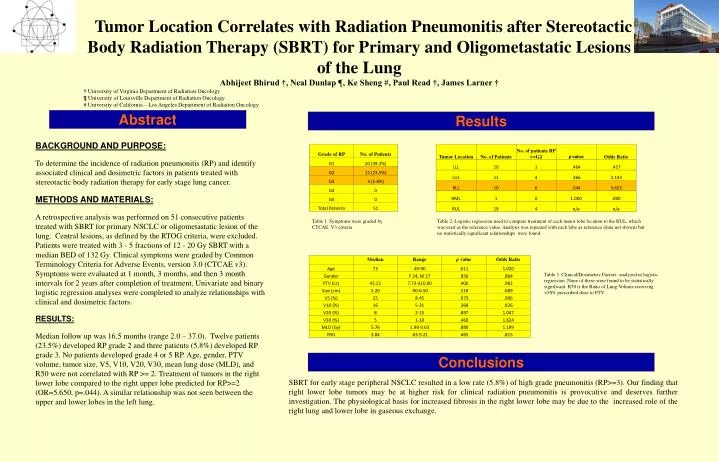
Tumor Location Correlates with Radiation Pneumonitis after Stereotactic Body Radiation Therapy (SBRT) for Primary and Oligometastatic Lesions of the Lung Abhijeet Bhirud †, Neal Dunlap ¶, Ke Sheng #, Paul Read †, James Larner † † University of Virginia Department of Radiation Oncology ¶ University of Louisville Department of Radiation Oncology # University of California – Los Angeles Department of Radiation Oncology Abstract Results BACKGROUND AND PURPOSE: To determine the incidence of radiation pneumonitis (RP) and identify associated clinical and dosimetric factors in patients treated with stereotactic body radiation therapy for early stage lung cancer. METHODS AND MATERIALS: A retrospective analysis was performed on 51 consecutive patients treated with SBRT for primary NSCLC or oligometastatic lesion of the lung. Central lesions, as defined by the RTOG criteria, were excluded. Patients were treated with 3 - 5 fractions of 12 - 20 Gy SBRT with a median BED of 132 Gy. Clinical symptoms were graded by Common Terminology Criteria for Adverse Events, version 3.0 (CTCAE v3). Symptoms were evaluated at 1 month, 3 months, and then 3 month intervals for 2 years after completion of treatment. Univariate and binary logistic regression analyses were completed to analyze relationships with clinical and dosimetric factors. RESULTS: Median follow up was 16.5 months (range 2.0 – 37.0). Twelve patients (23.5%) developed RP grade 2 and three patients (5.8%) developed RP grade 3. No patients developed grade 4 or 5 RP. Age, gender, PTV volume, tumor size, V5, V10, V20, V30, mean lung dose (MLD), and R50 were not correlated with RP >= 2. Treatment of tumors in the right lower lobe compared to the right upper lobe predicted for RP>=2 (OR=5.650, p=.044). A similar relationship was not seen between the upper and lower lobes in the left lung. Table 1. Symptoms were graded by CTCAE. V3 criteria Table 2. Logistic regression used to compare treatment of each tumor lobe location to the RUL, which was used as the reference value. Analysis was repeated with each lobe as reference (data not shown) but no statistically significant relationships were found. Table 3. Clinical/Dosimetric Factors analyzed in logistic regression. None of these were found to be statistically significant. R50 is the Ratio of Lung Volume receiving >50% prescribed dose to PTV. Conclusions SBRT for early stage peripheral NSCLC resulted in a low rate (5.8%) of high grade pneumonitis (RP>=3). Our finding that right lower lobe tumors may be at higher risk for clinical radiation pneumonitis is provocative and deserves further investigation. The physiological basis for increased fibrosis in the right lower lobe may be due to the increased role of the right lung and lower lobe in gaseous exchange.