Download
1 / 41
410 likes | 620 Views
Explore the intricacies of sexual reproduction, including meiosis stages, sperm production (spermatogenesis), and the male reproductive system anatomy. From germ cells to hormonal control, delve into the vital aspects of reproduction.

E N D
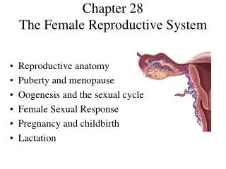
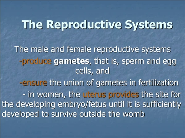
Chapter 28The Reproductive Systems • Sexual reproduction produces new individuals • germ cells called gametes (sperm & 2nd oocyte) • fertilization produces one cell with one set of chromosomes from each parent • Gonads produce gametes & secrete sex hormones • Reproductive systems • gonads, ducts, glands & supporting structures
Chromosomes in Somatic Cells & Gametes • Somatic cells (diploid cells) • 23 pairs of chromosomes for a total of 46 • each pair is homologous since contain similar genes in same order • one member of each pair is from each parent • 22 autosomes & 1 pair of sex chromosomes • sex chromosomes are either X or Y • females have two X chromosomes • males have an X and a smaller Y chromosome • Gametes (haploid cells) • single set of chromosomes for a total of 23 • produced by special type of division: meiosis
tetrad Meiosis I -- Prophase I • Chromosomes become visible, mitotic spindle appears, nuclear membrane & nucleoli disappear • Events not seen in prophase of Mitosis or Meiosis II • synapsis • all copies of homologous chromosomes pair off forming a tetrad • crossing-over • portions of chromatids are exchanged between any members of the tetrad • parts of maternal chromosomes may be exchanged with paternal ones • genetic recombination produces gametes unlike either parent
Exchange of Genetic Material • Chromosomes are exchanged between chromatids on homologous chromosomes
Meiosis I -- Metaphase I, Anaphase I & Telophase I • In metaphase I, homologous pairs of chromosomes line up along metaphase plate with attached microtubules • In anaphase I, each set of homologous chromatids held together by a centromere are pulled to opposite ends of the dividing cell • Telophase I and cytokinesis are similar to mitotic division • Result is 2 cells with haploid number of chromosomes
Meiosis II • Consists of 4 phases : prophase II, metaphase II, anaphase II and telophase II • Similar steps in this cellular process as in mitosis • centromeres split • sister chromatids separate and move toward opposite poles of the cell • Each of the daughter cells produced by meiosis I divides during meiosis II and the net result is 4 genetically unique haploid cells or gametes.
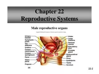
Male Reproductive System • Gonads, ducts, sex glands & supporting structures • Semen contains sperm plus glandular secretions
Scrotum • Sac of loose skin, fascia & smooth muscle divided into two pouches by septum • Skin contains dartos muscle causes wrinkling • Temperature regulation of testes • sperm survival requires 3 degrees lower temperature than core body temperature • cremaster muscle in spermatic cord • elevates testes on exposure to cold & during arousal • warmth reverses the process
Testes • Paired oval glands measuring 2 in. by 1in. • Surrounded by dense white capsule called tunica albuginea • septa form 200 - 300 compartments called lobules • Each is filled with 2 or 3 seminiferous tubules where sperm are formed
Sperm Morphology • Adapted for reaching & penetrating a secondary oocyte • Head contains DNA & acrosome (hyaluronidase and proteinase enzymes) • Midpiece contains mitochondria to form ATP • Tail is flagellum used for locomotion
Hormonal Control of Spermatogenesis • Puberty • hypothalamus increases its stimulation of anterior pituitary with releasing hormones • anterior pituitary increases secretion LH & FSH • LH stimulates Leydig cells to secrete testosterone • FSH stimulates spermatogenesis • with testosterone, stimulates sertoli cells to secrete androgen-binding protein (keeps hormones levels high) • testosterone stimulates final steps spermatogenesis
Pathway of Sperm Flow through the Ducts of the Testis • Seminiferous tubules • Straight tubules • Rete testis • Efferent ducts • Ductus epididymis • Ductus (vas) deferens
Epididymis • Comma-shaped organ, 1.5in long along posterior border of each testis • Head, body and tail region • Multiple efferent ducts become a single ductus epididymis in the head region • 20 foot tube if uncoiled • Tail region continues as ductus deferens
Epididymis • Site of sperm maturation • motility increases over 2 week period • Storage for 1-2 months • Propels sperm onward
Ductus (Vas) Deferens • Pathway of 18 inch muscular tube • ascends along posterior border of epididymis • passes up through spermatic cord and inguinal ligament • reaches posterior surface of urinary bladder • empties into prostatic urethra with seminal vesicle • Lined with pseudostratified columnar epithelium & covered with heavy coating of muscle • convey sperm along through peristaltic contractions • stored sperm remain viable for several months
Urethra • 8 inch long passageway for urine & semen • Prostatic urethra (1 inch long) • Membranous urethra (passes through UG diaphragm ) • Penile (spongy) urethra (through corpus spongiosum)
Seminal Vesicles • Pair of pouchlike organs found posterior to the base of bladder • Alkaline, viscous fluid • neutralizes vaginal acid & male urethra • fructose for ATP production • prostaglandins stimulate sperm motility & viability • clotting proteins for coagulation of semen Posterior View
Prostate Gland • Single organ the size of chestnut found inferior to bladder • Secretes milky, pH 6.5 fluid that increases sperm motility and viability • citric acid for ATP production & enzymes for seminal liquefaction • Many duct openings • Enlarges with age
Bulbourethral or Cowper’s Gland • Paired, pea-sized gland within the UG diaphragm • Secretes alkaline mucous into spongy urethra • Neutralizes acids and lubricates
Penis • Passageway for semen & urine • Body composed of three erectile tissue masses filled with blood sinuses • Composed of bulb, crura, body & glans penis
Glans Penis • Enlarged distal end of corpus spongiosum • External urethral orifice is small slit • Covered by loosely fitting prepuce or foreskin
Female Reproductive System • Ovaries produce 2nd oocytes & hormones • Uterine tubes transport fertilized ova • Uterus where fetal development occurs • Vagina & external genitalia constitute the vulva • Mammary glands produce milk
Follicular Stages • Stages of follicular development • primordial • primary • secondary • graafian • ovulation • Corpus luteum is ovulation wound • fills in with hormone secreting cells • Corpus albicans is white scar left after corpus luteum is not needed
Life History of Oogonia • Germ cells from yolk sac migrate to ovary & become oogonia • As a fetus, oogonia divide to produce millions by mitosis but most degenerate (atresia) • Some develop into primary oocytes & stop in prophase stage of meiosis I • 200,000 to 2 million present at birth • 40,000 remain at puberty but only 400 mature during a woman’s life • Each month, hormones cause meiosis I to resume in several follicles so that meiosis II is reached by ovulation • Penetration by the sperm causes the final stages of meiosis to occur
Uterine or Fallopian Tubes • Narrow, 4 inch tube extends from ovary to uterus • infundibulum is open, funnel-shaped portion near the ovary • fimbriae are moving finger-like processes • ampulla is central region of tube • isthmus is narrowest portion joins uterus
Histology & Function of Uterine Tube • Histology = 3 Layers • mucosa = ciliated columnar epithelium with secretory cells provide nutrients & cilia move along ovum • muscularis = circular & longitudinal smooth muscle • peristalsis helps move ovum down to the uterus • serosa = outer serous membrane • Function -- events occurring in the uterine tube • fimbriae sweep oocyte into tube, cilia & peristalsis move it along, sperm reaches oocyte in ampulla, fertilization occurs within 24 hours after ovulation & zygote reaches uterus about 7 days after ovulation
Anatomy of the Uterus • Site of menstruation& development of fetus • Description • 3 inches long by 2 in. wide and 1 in. thick • subdivided into fundus,body, isthmus & cervix • interiorly contains uterine cavity accessed by cervical canal (internal & external os)
Vagina • Passageway for birth, menstrual flow & intercourse • Description • 4 inch long fibromuscular organ ending at cervix • mucosal layer • stratified squamous epithelium & areolar connective tissue • large stores of glycogen breakdown to produce acidic pH • muscularis layer is smooth muscle allows considerable stretch • adventitia is loose connective tissue that binds it to other organs • lies between urinary bladder and rectum • orifice partially closed with membrane (hymen)
Vulva (pudendum) • Mons pubis -- fatty pad over the pubic symphysis • Labia majora & minora -- folds of skin encircling vestibule where find urethral and vaginal openings • Clitoris -- small mass of erectile tissue • Bulb of vestibule -- masses of erectile tissue just deep to the labia on either side of the vaginal orifice
Female Reproductive Cycle • Controlled by monthly hormone cycle of anterior pituitary, hypothalamus & ovary • Monthly cycle of changes in ovary and uterus • Ovarian cycle • changes in ovary during & after maturation of oocyte • Uterine cycle • preparation of uterus to receive fertilized ovum • if implantation does not occur, the stratum functionalis is shed during menstruation
Menstrual Phase • Menstruation lasts for 5 days • First day is considered beginning of 28 day cycle • In ovary • 20 follicles that began to develop 6 days before are now beginning to secrete estrogen • fluid is filling the antrum from granulosa cells • In uterus • declining levels of progesterone caused spiral arteries to constrict -- glandular tissue dies • stratum functionalis layer is sloughed off along with 50 to 150 ml of blood
Preovulatory Phase • Lasts from day 6 to 13 (most variable timeline) • In the ovary (follicular phase) • follicular secretion of estrogen & inhibin has slowed the secretion of FSH • dominant follicles survives to day 6 • by day 14, graafian follicle has enlarged & bulges at surface • increasing estrogen levels trigger the secretion of LH • In the uterus (proliferative phase) • increasing estrogen levels have repaired & thickened the stratum functionalis to 4-10 mm in thickness
Ovulation • Rupture of follicle & release of 2nd oocyte on day 14 • Cause • increasing levels of estrogen stimulate release of GnRH which stimulates anterior pituitary to release more LH • Corpus hemorrhagicum results
Signs of Ovulation • Increase in basal body temperature • Changes in cervical mucus • Cervix softens • Mittelschmerz---pain
Postovulatory Phase • Most constant timeline = lasts 14 days • In the ovary (luteal phase) • if fertilization did not occur, corpus albicans is formed • as hormone levels drop, secretion of GnRH, FSH & LH rise • if fertilization did occur, developing embryo secretes human chorionic gonadotropin (hCG) which maintains health of corpus luteum & its hormone secretions • In the uterus (secretory phase) • hormones from corpus luteum promote thickening of endometrium to 12-18 mm • formation of more endometrial glands & vascularization • if no fertilization occurs, menstrual phase will begin
Homework: Chapter • B5, B8, B10, B12, B13, C1, C14