Download
1 / 53
550 likes | 781 Views
Neural crest and placods . Krisztina H.-Minkó PhD Deptartment of Anatomy, Histology and Embryology Semmelweis University, Faculty of Medicine. 2018. 3 -week old human embryo. Neural crest.

E N D
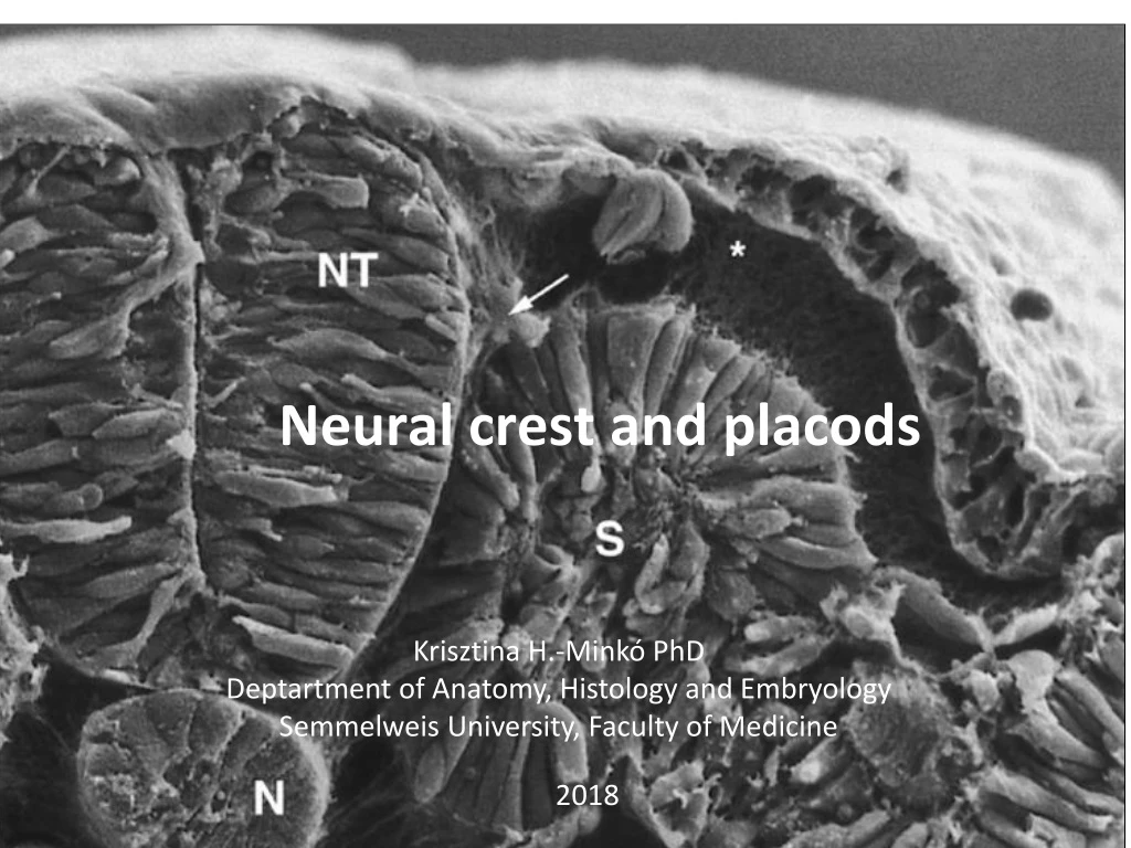
Neural crest and placods Krisztina H.-Minkó PhD Deptartment of Anatomy, Histology and Embryology Semmelweis University, Faculty of Medicine 2018
Neural crest Figure 12-2 Scanning electron micrograph of a chick embryo, showing the early migration of neural crest cells (arrow) out of the neural tube (NT). The subectodermal pathway of neural crest migration (*) is relatively cell-free, but contains a fine mesh of extracellular matrix molecules. N, notochord; S, somite. (Courtesy K. Tosney, Ann Arbor, Mich.)
Neural tube closure: http://www.youtube.com/watch?v=ZcAAsr_8vOE Migrationof NC cells in the head of chiken embryo: http://www.youtube.com/watch?v=IP0IsQ5QYgo
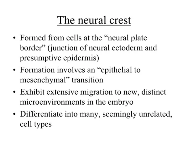
Neural crest specification at the border of neural and epidermal ectoderm
Prof. Csillag Early differentiation and partitioning of neural plate
Placodes and neural crest Neural crest: caudal, medial, epithelio-mesenchymal transition Placode: cranial, lateral, epithelialial tickenings may form vesicles Prof. Szél
Placode Derivative Hypophyseal Olfactory Lens Trigeminal Otic Epibranchial Neural plate Neural crest Ectoderm Rathke’s pouch Adenohypophysis Olfactory epithelium Lens Trigeminal ggl. (partly) Otic vesicle membranous labyrinth, spiral+vestibular ggl.* (VIII.) Epibranchial –> taste buds, geniculate ggl. (VII.), inf. ggl. of IX. and X. nerves* Brain, spinal cord ganglia, … epidermis of skin,… *Special sensory ganglia!
Neural crest and the placodes, two transient ectodermal cell populations in the embryo Neural crest cells give advantages to Vertebrates Most of the morphological and functional differences between vertebrates and other chordates occur in the head and are derived embryologically from muscularized hypomere, neural crest, and epidermal (neurogenic) placodes. In the head, the neural crest functions as mesoderm and forms connective, skeletal, and muscular tissue. Both the neural crest and the epidermal placodes form special sense organs and other neural structures. These structures may be homologous to portions of the epidermal nerve plexus of protochordates. The transition to vertebrates apparently was associated with a shift from a passive to an active mode of predation, so that many of the features occurring only in vertebrates became concentrated in the head.(Gans és Northcutt, Science 1983) Plasticity of neural crest cells… ecto-mesenchyme Characterictics of the vertebrate „New Head”: Special composite sense organs, complex visual organ is important in predation Jaws for predation! Complex viscerocranium and chondrocranium development (Neuron. 2003 Mar 27;37(6):895-8.A celebration of the new head and an evaluation of the new mouth.)
Twenty years ago now, Carl Gans and Glen Northcutt proposed that the main invention of vertebrates was a new head, with its full array of sensory organs involved in an active predatory lifestyle. Tracing back the embryological origin of these structures, they showed how all are primarily derived from the neural crest and the placodes, two transient ectodermal cell populations in the embryo. These cell types were then used for further innovations, such as a new mouth in jawed vertebrates. The interplay between patterning and plasticity of the neural crest is largely responsible for the endless variation of vertebrate craniofacial features in evolution. (Neuron. 2003 Mar 27;37(6):895-8.A celebration of the new head and an evaluation of the new mouth.) The appearance of the neural crest and theplacodes provided the basis for the formation of pairedsense organs and structures at the anterior part of thehead, which represented a new vertebrate unit and allowed an active predatory lifestyle. These new cellulartypes gave rise to new structures that are characteristic of the vertebrate head. More advantages: Development of the peripheral nervous system Pigment cells against UV radiation, accomodation to environment
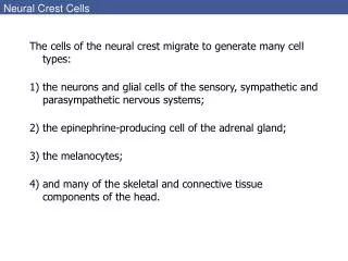
Derivatives of the neural crest Ganglia, peripheral nerve cells glial cells Myofibroblast, fibroblast Cartilage, bone melanocytes endocrine cells
First discovery by His in the chiken embryo (1868). Mediodorsal cells immigrate laterally to invade spinal ganglia He named them „ganglionic crest” cells. Further extirpation experiments showed that not only sensory but sympathetic and parasympathetic ganglia and enteric nervous system derive from the neural crest
Quail-chick chimera fate mapping studies 2. heterochromatin is different in quail nuclei, lot of heterochromatin associated to the nucleolus dispersal heterokromatin in chicken nuclei
Quail-chick chimera fate mapping studies 3. NCC: neural crest cells
Neural crest specification: migrating into genomics Laura S. Gammill & Marianne Bronner-Fraser Nature Reviews Neuroscience4, 795-805 (October 2003) | doi:10.1038/nrn1219
Results of chimera experiments, fate mapping studies - derivatives of the neural crest enteric nervous system adrenal medulla N.M. Le Douarin / Mechanisms of Development 121 (2004) 1089–1102
Derivatives • peripheral nervous system • sympathetic, parasympathetic, sensory, autonome • Schwann cells • melanocytes • cartilage and bone in the head, smooth muscle, myofibroblast and fibroblast , mesectoderm • endocrine cells (adrenal medulla) • Meninges (pia, arachnoid) • vessel wall (not endothelia) in the head region • Heart : aortico-pulmonal, conotruncal septum New head and heart story
Neural crest induction, current modell Signals from ectoderm: BMP, Wnt from mesoderm FGF-8 (amphibian data) high conc. of BMP: epidermal ectoderm, low: neural ectodermmedium BMP conc. defines neural crest Msx-1, Pax-3 expression starts (characteristic for NC cells) snail-1 és slug (snail-2) expression starts which is neede for EMT(epithelio-mesenchymal transition) slug is expressed at gastrulation as well! Changes in adhesion properties (adhesion molecules) downregulation of N-CAM, E-cadherin, and N-cadherin, and re.upregulation with integration into tissues, cadherin 7 and 11 expr. upregulated
After induction: migration In the head region neural crest cells start the migration before the closure of the neural tube, not in the trunk Fibronectin, laminin, and type IV. collagen are favorable chondroitin-sulfate: non-favorable cell-surface integrins In the head region neural crest cells start the migration before the closure of the neural tube, not in the trunk HEAD TRUNK
Neural crest divisions Circumpharyngeal pharynx heart, great vessels intestine trunk 6th somite sacral cranial
Cranial craniofacial (ecto-) mesenchyme, cartilage, bone, conn. tissue, nerve, glia Topography Cardiac arterial wall, septum aorticopulmonale Enteric vagal, sacral, parasympathetic elements Trunk melanocytes, spinal ganglia, sympathetic ganglia Prof. Szél
Neuromeres, prosomeres, rhombomeres http://www.cram.com/flashcards/neuro-47-development-of-the-nervous-system-2544719
5th and 6th prosomere level do not give rise to neural crest only 1-4 prosomeres. NC from hindbrain levels colonise 1st-3rd pharyngeal arches. Each rhombomeric and mesencephalic crest cell „remember” to the segmental code. In the pharyngeal region, the pathways of crest cell migration are cosely correlated with Hoxb gene expr. Cells of the cranial crest may be patterned with level –specific instructions, whereas cells of the trunk crest are not.
NC cell migration in 3 streams: • ‘trigeminal’ – around 3 divisions of CN V. (1st branchial arch*+frontonasal process) • ‘hyoid’ – into 2. branchial arch • ‘postotic’ (= behind otic vesicle) – into branchial arches 3-6. Ectomesenchymw: bones, cartilage, conn. tissue, vessels * 2 of 3 auditory ossicles, jaw (Meckel’s cartilage)- skull… „new head” Rhombomeres 1,2,(3) Rhombomeres (3),4,(5) Rhombomeres (5),6,7,8
N.M. Le Douarin / Mechanisms of Development 121 (2004) 1089–1102 • Hox genes determines segments and NC cells do not mix between these segments
DiGeorge syndrome - hoxa-3 gene defect Mesenchymal elements of cranialis crest (III-IV. pharyngeal arches) are defective Aplasia of thymus and parathyroid gland (III-IV. pharyngeal pouch), „fish-mouth” deforation (shorter philtrum), deformation of lingual and cervical muscles, hypertelorism, mal-formations of the heart Knockout mouse: short, thicke neck, lack of thymus and parathyroid gland, deformation of cardiac vessels and valves, deformation of greater horn of hyoid bone, cricoid cartilage, and epiglottis Prof. Szél
The circumpharyngeal neural crest arises in the posterior rhombencephalic region, and in the lower part of the pharynx, emigrating circumpharyngeal crest cells pass behind the sixth pharyngeal arch. Neural crest cells from the anterior rhombencephalon to the level of somite 5 emigrate from the circumpharyngeal crest as a stream, called the cardiac crest, toward the developing heart and aortic arches, whereas other neural crest cells from the levels of somites 1 to 7 constitute the vagal crest and migrate into the developing gut as precursors of the parasympathetic innervation of the digestive tract.
19-day old chicken embryo colon Remak ganglion plexus myentericus plexus submucosus Goldstein and Nagy, 2008, vol 64. Ped Res
Trunk neural crest Migratory pathways ventrolateral pathway (anterior somite: sensory ganglia) dorsolateral pathway (melanocytes) ventral pathway (sympathico-adrenal) Prof. Szél
Migration in the trunk • BMP4 • Ventromedial pathway: • rostral part of somite • ephrin B1 caudally, non favorable posterior sclerotom inhibite migrtion because of the presence of semaforins and condroitin-sulfate • sensory and sympathetic ganglia, Schwann and chromaffin cells • In accordance with somite development and compartmentalisation • Dorsolateral pathway: • between somites and ectoderm, no segmentation • 24 hours after the beginning of ventromedial migration • melanocytes; endothelin-3 • glicoprotein expression regulates which patway to choose • ECM: tenascin, thrombospondin –activating, F-spondin, collagen IX – inhibiting • perinotochordal mesenchyme (chondroitin-sulfate) dermomyotome sclerotom Krull (2001) MOD, 105:37-45.