Download
1 / 49
550 likes | 970 Views
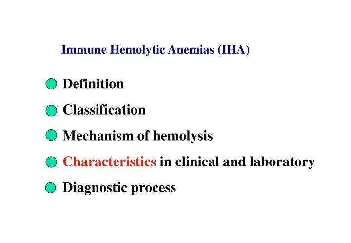
Definition Classification Mechanism of hemolysis Characteristics in clinical and laboratory Diagnostic process. Immune Hemolytic Anemias (IHA). Definition of IHA

E N D
Definition Classification Mechanism of hemolysis Characteristics in clinical and laboratory Diagnostic process Immune Hemolytic Anemias (IHA)
Definition of IHA The shortened red cells life span is caused by the antigens existing on the surface of erythrocytes reacting with relative antibodies. This acquired hemolytic anemia is termed immunehemolytic anemia.
Classification 1. According to the cause of Ab production 2. According to the nature of Ab temperature Autoimmunehemolytic anemia (AIHA) Drug-induced immune hemolytic anemia (DIHA) Allo-immune hemolytic anemia (ALIHA) Warm Ab type (37 ℃,IgG) Cold Ab type (less than 37 ℃, IgM) Mixed Ab type IHA
I. Autoimmunehemolytic anemias (AIHAs) • Definition AIHAs are a group of acquired hemolytic disorders that are the result of antibodiesor complement binding to specific antigens on the RBC membrane, which leads to ashortened RBC life span and an increased RBC destruction.
Antierythrocyte antibodies can be divided into three general categories: 1. IgG Warm Abs bind to RBCs at 37 ℃ but fail to agglutinate the RBCs. 2. Cold agglutinins almost always are of IgM subtype and clump RBCs at cold tempratures. (CAS) . 3. Donath-Landsteiner(IgG) Abs bind to RBCs in the cold and activate the hemolytic complement cascade when the RBCs are warmed to 37 ℃. (PCH)
overview: • a group of diseases resulting from the binding of IgG to the RBC membrane. • The IgG-bound RBCs either are trapped by macrophages as they pass through the spleen or may first have part of their membrane removed, then destroyed in the circulation. • 70% of AIHAs. AIHA : Warm Antibody Type
Pathogenesis of hemolysis: 37°C IgG macrophages FcR Spleen sinus C3bR spleen extravascularand/or intravascular
Etiology: • Idiopathic: females more than males? • secondary causes: • Lymphoprolitive diseases(e.g., CLL , non-Hodgkin lymphoma ) • Connective tissue diseases(e.g., SLE) • Immune deficiency disorders( e.g., AIDS) • Drugs(e.g., penicillin, quinidine, methyldopa, cephalosporins)
Clinical Features 1. jaundice, anemia(e.g., fatigue, shortness of breath, light-headedness) or asymptomatic 2 . PE: jaundice, splenomegaly, and some signs relate to underlying illness , such as fever, lymphadenopathy, skin rash,, hypertension, renal failure, petechiae or ecchymoses ( PC decrease) Evans syndrome is the combination of warm AIHA and idiopathic thrombocytopenia purpura. Highly variable symptoms that are unrelated to temperature.
Lab Findings Blood: Hb, PCV , RBC( vary from normal to low) Ret , “3M” (MCV ?) and RDW fall Morphology:polychromatic RBCs, macrocytosis, nucleated RBCs, or microspherocytes. WBC? PC: N or low Bone marrow: hyperplasia figure Serum bilirubin and urobilinogen(urine):elevated DAT usually positive. ( confirm test)
Bone marrow reveals erythroid hyperplasia. Polychromatic and orthochromatic normoblasts increaes, M/E decreased, erythroid mitosis increased; mature RBC?
serum Anti-immunoglobin Poly- Coombs Test 1. Direct Anti-immunoglobin Test ( DAT ) Anti-IgG Anti-complement
Cold AIHA • Cold Agglutinin Syndrome (CAS) overview • a group of disorders caused by IgM auto-antibodies (usually against the I/i antigen) • cold agglutinins bind to RBCs at cold temperatures (4 ℃ - 18 ℃).
cold agglutinins-IgM binding at low temperature activate complement, and C3b fixation( at higher tempratures.) 0 to 5 (4~18) ℃ anti-C3+ agglutinate Hemolytic mechanism of CAS : Extravascular or Intravascular
Specific clinical features of CAS 1.Cold-induced acrocyanosis (Raynaud’s phenomenon): blue color of the skin at fingertips, toes, nose, and ear lobes) due to vascular sludding arising from agglutination. 2. cold-associated hemoglobinuria 3. Secondary to upper respiratory tract infection 4. Symptoms worse in cold weather and higher IgM titer and activity)
Lab Findings: --anemia: Hb --Hemolysis: ret, bilirubin, LDH, --blood smear: RBC agglutination --bone marrow --DAT +: for C3 only. (screening) --The Cold agglutinin test(confirm)
serum Anti-immunoglobin Poly- Anti –C3 + Coombs Test Direct Anti-immunoglobin Test ( DAT )
4℃ to 30℃ 37℃ autoerythrocytes or O type RBC or the same type RBC Patient serum cold agglutinin IgM Cold agglutinin test
paroxysms of fever, back pain, leg pain, • abdominal cramps; rigors (after exposure to cold temp.) hemoglobinuria • hemolysis: intravascular , less than 20 ℃ • children often , secondary to viral disorder syphilis III. Paroxysmal cold hemoglobinuria (PCH) • IgG autoantibodies are specific for the P antigen. • D-L Ab: mono- or polyclonal; IgG;
Lab findings: 1. Hb, PCV (severity of anemia) 2. Ret low in episode; elevated in recovery phase. 3. Blood smear: anisocytosis, poikilocytosis, polychromatophilia, spherocytes, nucleated RBCs. 4. FHb , Hp ; hemoglobinuria, methemoglobin (urin) 5 DAT negative usually, or C3 + only, IAT+ 6 Donath-Landstainer Test : D-L Ab +
Sample of patient T<20 ℃ Anti-IgG,IgM or / and C Indirect Anti-immunoglobin Test ( IAT )
Cold Warm Hemolysis Test (Donath-Landstainer Test) 【Principle】 At lower temperature(<20℃), D-L antibody may attach to the surface of the patient’s red cell. When temperature up to 37℃, the red cells with D-L antibodies lysis rapidly with the help of activated complements. 【Result】 Negative: no lysis in normal people.
<20℃ 37℃
Hb, RBC, PCV; 3M, RDW Fatigue, weakness Anemia and type of anemia jaundice, hemoglobinuria Splenomegaly, fever, chilly Ret morphology serum and urine Hemolytic anemia and its type underlying diseases and specific featrues? IHA Coombs test Anti-C3 Anti-C3, ITA+ DAT+ cold agglutinin test cold warm hemolysis test titer>1:64 Warm type of AIHA PCH PLC CAS Evan syndrome
Questions : • Summarize the features of blood and bone marrow smear in IHA? • How do you know the patient may have IHA?
Case A female, 45 years old Complaintof “having been fatigue, weakness for about 6 months. Sometimes had black urine.” PE: moderate splenomegaly and slightly enlarged liver. Lab: Hg 80g/l, Hct 0.30 , RBC 2.5X10¹² , RDW 16.7 What do you know from these information?
1 2 3 4 5 No.5 is the result of Coombs test of the patient DAT(anti IgG+anti C3)
Discussing questions: 1. What is your primitive impression of the patient? Describe your evidence? 2. What clinical information do you need to support your diagnosis? 3. Which further tests do you need to complete the diagnosis? 4. Why the patient had black urine.
Secondary anemia MYELOPATHIC ANEMIAS These anemias are due to the bone marrow infiltration or replacement by abnormal tissues such as malignant metastases, myelosclerosis, leukemia and myeloma.
Pathogenesis of myelopathic anemia: --bone marrow damage --hemorrhage --hemolysis
Clinical Features --severe anemia --bleeding problems may occur --splenomegaly and hepatomegaly is common --symptoms referable to underlying disease
Lab Findings Blood --varying degrees of anemia (most normocytic; slightly macrocytic) --distinct anisocytosis and poikilocytosis --leukoerythroblastic anemia: --reticulocytosis and polychromatophilia --The WBC count may be variable --The platelet count is often low, giant, bizarre shaped with abnormal function.
disruption of marrow sinusoids hematopoiesis in extramedullary sites Why? What isleukoerythroblastic anemia? The patients suffering from anemias with nucleated RBCs and immature granulocytes in the peripheral blood.
.Bone marrow --The marrow aspiration may fail or show metastatic cellsand so on. --The marrow biopsy is necessary to establish the diagnosis. Other tests: --x-ray --NAP --CD
ANEMIA REVIEW
贫 血 急性失血性贫血 慢性失血性贫血 微血管病性溶血性贫血 化学、物理、生物因素致溶血 脾功能亢进 阵发性睡眠性血红蛋白尿症 遗传性椭圆红细胞增多症等 遗传性球形红细胞增多症 葡萄糖6磷酸脱氢酶缺乏症 丙酮酸激酶缺乏症等 珠蛋白生成障碍性贫血 异常血红蛋白病 不稳定血红蛋白病 肾病、肝病、感染性疾病、 内分泌疾病等 缺铁性贫血 铁粒幼细胞性贫血等 白血病、骨髓瘤、癌转移、 骨髓纤维化等 再障,纯红再障等 骨髓增生异常综合征等 红细胞生成减少 红细胞破坏过多 红细胞丢失增加 红细胞 内在缺陷 外在异常 骨髓造血功能障碍 造血物质缺乏或利用障碍 膜异常 酶异常 Hb异常 免疫因素 非免疫因素 干细胞增殖分化障碍 骨髓被异常组织侵害 骨髓造血功能低下 铁缺乏和铁利用障碍 维生素B12或叶酸缺乏 各种原因致免疫性 溶血性贫血 巨幼细胞贫血等 贫血的病因及发病机制分类
Anemia: weakness,fatigue, listlessness,palpitation, pallor jaundice, splenomegaly MCV increase normal decrease MCH MA IDA,SA,Thala MCHC infection decrease ret increase acute loss blood hypopoiesis HA (Coomb’s) decrease PL normal IHA infection extracellular defect intra- WBC chronic renal disease osmotic fragility decrease increase increase normal decrease AA,MF AM HS,HE, G-6PD, PK Thala PNH (ham’s) abnormalHb
Diagnostic steps of HA: history,infection, underlying diseases,drugs,pallor,weakness dark urine, jaundice,hepatosplenomegaly blood film spherocytes, autoagglutination, red cell fragments ret increased immune assay cold agglutinin test Coomb’s test positive CAS AIHA negative cold warm hemolysis test transfusion reaction infection, congenital syphilis
Normocytic normochromic anemia history, underlying diseases, pallor,weakness blood film decrease/ increase ret increase morphologic features acute blood loss HA normal abnormal secondary hypoplasia abnormal proliferation anemia infiltration in marrow Infection AA MDS leukemia renal disease MF liver disease metastatic cancer endocrinic disease
Microcytic hypochromic anemia: history, underlying diseases, anemia,MCV,MCH Blood film SI increased normal/ increased decreased Iron stain Hb eletrophoresis SF HbA2,F increased normal/ increased decreased SA Thalasemia anemia of chronic IDA HbC,S, D,E disorders etc.
Differentiation of macrocytic anemias:MCV, MCH history, anemia ,underlying diseases, drugs,nutrition,neurologic signs, hepatosplenomegly blood film increased Ret normal / decreased marrow morphology acute blood loss HA non-MA MA erythroblastic anemia abnormal proliferation Alchohol poisoning MDS Folate deficiency Liver disease VitB12 deficiency Pernicious anemia