Download
1 / 1
10 likes | 118 Views
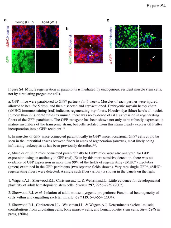
Figure S4. Young (GFP) Aged (WT). GFP eMHC / Hoechst. -GFP eMHC / Hoechst. a. c. b. GFP / eMHC / Hoechst. Figure S4 Muscle regeneration in parabionts is mediated by endogenous, resident muscle stem cells, not by circulating progenitor cells.

E N D
Figure S4 Young (GFP) Aged (WT) GFP eMHC/Hoechst -GFP eMHC/Hoechst a c b GFP/eMHC/Hoechst • Figure S4 Muscle regeneration in parabionts is mediated by endogenous, resident muscle stem cells, not by circulating progenitor cells. • a, GFP- mice were parabiosed to GFP+ partners for 5 weeks. Muscles of each partner were injured, allowed to heal for 5 days, and then dissected and cryosectioned. Embryonic myosin heavy chain (eMHC) immunostaining (red) indicates regenerating myofibers. Hoechst dye (blue) labels all nuclei. In more than 99% of the fields examined, there was no evidence of GFP expression in regenerating fibers of the GFP- parabionts. The GFP transgene has been shown not only to be robustly expressed in mature myofibers of the transgenic strain, but cells isolated from this strain clearly express GFP after incorporation into a GFP- recipient1-3. • b, In muscles of GFP- mice connected parabiotically to GFP+ mice, occasional GFP+ cells could be seen in the interstitial spaces between fibers in areas of regeneration (arrows), most likely being infiltrating leukocytes as has been previously described1,3. • c, Muscles of GFP- mice connected parabiotically to GFP+ mice were also analyzed for GFP expression using an antibody to GFP (red). Even by this more sensitive detection, there was no evidence of GFP expression in more than 99% of the fields of regenerating (eMHC+) myotubes (green) examined in the GFP- parabionts (two separate fields shown). Very rare single GFP+, eMHC+ regenerating fibers were detected. A single such fiber (arrow) is shown in the panels on the right. • 1. Wagers,A.J., Sherwood,R.I., Christensen,J.L. & Weissman,I.L. Little evidence for developmental plasticity of adult hematopoietic stem cells. Science297, 2256-2259 (2002). • 2. Sherwood,R.I. et al. Isolation of adult mouse myogenic progenitors: Functional heterogeneity of cells within and engrafting skeletal muscle. Cell119, 543-554 (2004). • 3. Sherwood,R.I., Christensen,J.L., Weissman,I.L. & Wagers,A.J. Determinants skeletal muscle contributions from circulating cells, bone marrow cells, and hematopoietic stem cells. Stem Cells in press, (2004).