Download
1 / 79
800 likes | 829 Views
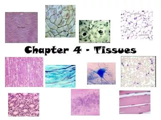
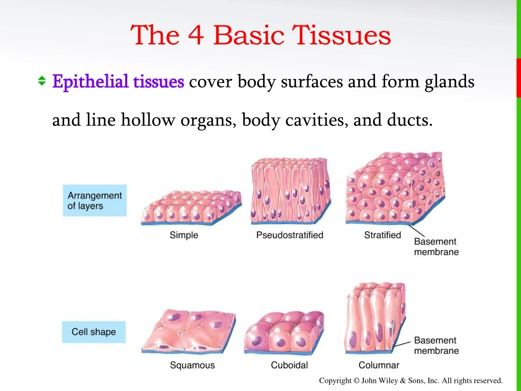
The 4 Basic Tissues. Epithelial tissues cover body surfaces and form glands and line hollow organs, body cavities, and ducts. Epithelium. Epithelium is used to line surfaces and form protective barriers. Epithelium is also good at secreting things like mucous, hormones, and

E N D
The 4 Basic Tissues • Epithelial tissues cover body surfaces and form glands and line hollow organs, body cavities, and ducts.
Epithelium • Epithelium is used to line surfaces and form protective barriers. Epithelium is also good at secreting things like mucous, hormones, and other substances . • All epithelia have a free apical surface and an attached basal surface.
Epithelium • The basal layer of the epithelium secretes a basal lamina; the underlying C.T. secretes a reticular lamina. • Together the basal lamina and the reticular lamina form a non- cellular basement membrane on which the epithelium sits.
Epithelium • Epithelia are named according to the shape of their cells, and the thickness or arrangement of their layers (of cells).
Epithelium • Naming epithelia according to shape
Epithelium • Naming epithelia according to arrangement
Epithelium • Naming epithelia • Three different cell shapes x three different cell arrangements = nine possibilities. Two of these are not used. Add transitional (cells that change shape), and we’re back up to eight possible combinations. • If different shapes are present in layers of cells, the epithelium is always named by the shape of cells in the apical (outermost) layer.
Epithelium • Simple Squamous Epithelium is composed of a single layer of flat cells found: • In the air sacs of lungs • In the lining of blood vessels, the heart, and lymphatic vessels • In all capillaries, including those of the kidney • As the major part of a serous membrane
Epithelium • Simple Cuboidal Epithelium is composed of a single layer of cube shaped cells. • It is often found lining the tubules of the kidneys and many other glands.
Epithelium • Simple Columnar Epithelium forms a single layer of column-like cells, ± cilia, ± microvilli, ± mucous (goblet cells). • Goblet cells are simple columnar cells that have differentiated to acquire the ability to secrete mucous.
Epithelium • Pseudostratified Columnar Epithelium appears to have layers, due to nuclei which are at various depths. In reality, all cells are attached to the basement membrane in a single layer, but some do not extend to the apical surface. • Ciliated tissue has goblet cells that secrete mucous.
Epithelium • Stratified Squamous Epithelium has an apical surface that is made up of squamous (flat) cells. • The other layers have different shapes, but the name is based on the apical layer. • The many layers are ideal for protection against strong friction forces.
Epithelium • Stratified Cuboidal Epithelium has an apical surface made up of two or more layers of cube-shaped cells. • Locations include the sweat glands and part of the ♂ urethra • Stratified Columnar Epithelium is very rare, and for our purposes, hardly worth mentioning.
Epithelium • The cells of Transitional Epithelium change shape depending on the state of stretch in the tissue. • The apical “dome cells” of the top layer (seen here in relaxation) are an identifiable feature and signify an empty bladder . • In a full bladder, the cells are flattened.
Epithelium • Although epithelia are found throughout the body, certain ones are associated with specific body locations. • Stratified squamous epithelium is a prominent feature of the outer layers of the skin.
Epithelium • Simple squamous makes up epithelial membranes and lines the blood vessels. • Columnar is common in the digestive tract. • Pseudostratified ciliated columnar is characteristic of the upper respiratory tract. • Transitional is found in the bladder. • Cuboidal lines ducts and sweat glands.
Covering and Lining Epithelium • Endothelium is a specialized simple squamous epithelium that lines the entire circulatory system from the heart to the smallest capillary – it is extremely important in reducing turbulence of flow of blood. • Mesothelium is found in serous membranes such as the pericardium, pleura, and peritoneum. • Unlike other epithelial tissue, both are derived from embryonic mesoderm (the middle layer of the 3 primary germ layers of the embryo).
Epithelial Membranes • Combining two tissues creates an organ. However, most of the organs and all of the organs systems studied this year contain all 4 basic types of tissues. • Epithelial membranes are the simplest organs in the body, constructed of only epithelium and a little bit of connective tissue.
Epithelial Membranes • Epithelial membranes = epithelium + connective tissue • Mucous membranes • Serous membranes • Cutaneous membrane = skin • Skin is not a simple organ. We will study the integument as our first organ system in the next chapter.
Epithelial Membranes • Mucous membranes line “interior” body surfaces open to the outside: • Digestive tract • Respiratory tract • Reproductive tract • Serous membranes line some internal surfaces: • Parietal layer next to body wall • Serous fluid between layers • Visceral layer next to organ
Epithelial Membranes • Skin as a cutaneous membrane is studied in Chapter 5.
Synovial Membranes • Synovial membranes enclose certain joints and are made of connective tissue only.
Glands • Epithelial glands are another example of simple organs • Glands that secrete their contents directly into the blood are called endocrine glands. • Glands that secrete their contents into a lumen or duct are called exocrine glands. • We will look at some common types of exocrine glands (endocrine glands are studied in Chapter 18.)
Exocrine Glands • Exocrine glands secrete substances through ducts to the surface of the skin or into the lumen of a hollow organ. • Secretions of the exocrine gland include mucus, sweat, oil, earwax, saliva, and digestive enzymes. • Examples of exocrine glands are sudoriferous (sweat) glands.
Exocrine Glands • Apocrine glands “bud” their secretions off through the plasma membrane, producing membrane-bound vesicles in the lumen of the gland. • The end of the cell breaks off by “decapitation”, leaving a milky, viscous odorless fluid. • This type of sweat only develops a strong odor when it comes into contact with bacteria on the skin surface.
Chapter 5 The Integumentary System Lecture slides prepared by Curtis DeFriez, Weber State University
Introduction • The organs of the integumentary system include the skin and its accessory structures including hair, nails, and glands, as well as blood vessels, muscles and nerves. • Note that all 4 of the basic tissue types are well-represented in this organ system: Epithelium in the hair, nails, and the epidermis of the skin; the dermis contains C.T.; muscle is found attached to the hair follicles, and in the substance of arteries and veins; nerves provide an abundance of sensation.
Introduction • The Integument is an organ system comprised of many organs such as hair and multiple types of glands.
Introduction • The integument can also be thought of as a cutaneous membrane that covers the outer surface of the body. • It is the largest organ by surface area and weight. • Its area is about 2 square meters (22 square feet) and weighs 4.5–5kg (10–11 lb), about 16% of body weight. • It is 0.5–4 mm thick, thinnest on the eyelids, thickest on the heels. • We lose almost a kg of skin epithelium a year that becomes a major part of household “dust”.
Introduction • Besides protection, the skin contributes to: • Regulation of body temperature • Sensory perceptions • Synthesis of vitamin D • Absorption and excretion • It also serves as an important reservoir of blood.
Structures of the Skin • The skin has 3 major layers: • The outer, thinner layer is called the epidermis and consists of epithelial tissue. • The inner, thicker layer is called the dermis and consists of C.T. • The subcutaneous (subQ) layer (also called the hypodermis) is located underneath the dermis. • It is a loose areolar/adipose C.T. that attaches the skin to the underlying tissues and organs.
Structures of the Skin • Dermatologist are doctors who treat disorders of all layers of the integumentary system.
The Epidermis • The epidermis is composed of keratinized stratified squamous epithelium which contains four major types of cells: • Keratinocytes • Melanocytes • Langerhans cells • Merkel cells
The Epidermis • Keratinocytes make up 90% of the cells. They produce keratin - a tough fibrous protein that provides protection. • Melanocytes produce the pigment melanin that protects against damage by ultraviolet radiation. • Langerhans cells are macrophages that originated in the red bone marrow. They are involved in the immune responses. • Merkel cells function in the sensation of touch along with the other adjacent tactile discs (receptors).
The Epidermis • The epidermis is composed of four layers in thin skin, and five layers in thick skin. • The stratum basale or stratum germinativum is always the bottom (deepest) layer. Continuous cell division occurs here and produces all the other layers. • The stratum spinosum is a layer of 8–10 keratinocytes • The non-dividing cells of the 3rd layer (stratum granulosum) are filled with granules of keratin.
The Epidermis • The stratum lucidum is the 4th layer but is only present in thick skin (the skin of the fingertips, palms, and soles). • The stratum corneum is always outermost, composed of approximately 20 layers of flat cell-remnants that are like “bags of turtle wax” (dead keratinocytes with no cellular organelles filled with only keratin protein.) • They are continuously shed and replaced by cells from deeper strata.
The Epidermis • Keratinization is the process of replacing viable cells in the stratum basale with more and more of the waxy keratin protein as cells move from the deepest layer to the surface layer. • Constant friction can stimulate the process and produce a callus (an even thicker buildup of keratinocytes in the stratum corneum). • Dandruff is an excess of keratinized cells shed from the scalp.
The Epidermis • Types of skin: • Thin (hairy) skin covers all body regions except the palms, palmar surfaces of digits, and soles. • Thick (hairless) skin covers the palms, palmar surfaces of digits, and soles.
The Epidermis • Skin Pigments • Melanin is produced by melanocytes in the stratum basale • Eumelanin (brown to black) • Pheomelanin (yellow to red) • Freckles are clusters of concentrated melanin triggered by exposure to sunlight. • Having more freckles is a genetic trait.
The Epidermis • Skin Pigments • Nevi (“birthmarks” or moles) are chronic lesions of the skin – they are, by definition, benign. • Malignant melanoma is a cancer of melanocytes.
The Epidermis • Skin Pigments • Vitiligo is a chronic disorder that causes depigmentation patches in the skin. The precise pathogenesis, or cause, is not known, but is most likely a combination of genetic factors coupled with a disorder of the immune system (autoimmune disease).
The Epidermis • Skin Pigments • Albinism is a congenital disorder characterized by the complete or partial absence of pigment in the skin, hair, and eyes due to a defect of an enzyme involved in the production of melanin.
The Dermis • The dermis is composed of connective tissue containing collagen and elastic fibers. • It contains two regions: • The papillary region lies just below the epidermis and consists of areolar connective tissue containing thin collagen and elastic fibers, dermal papillae (including capillary loops), corpuscles of touch and free nerve endings.
The Dermis • The reticular region consists of dense irregular connective tissue containing collagen and elastic fibers, adipose cells, hair follicles, nerves, sebaceous (oil) glands, and sudoriferous (sweat) glands. • Tears or excessive stretching in this region cause stretch marks (also called striae).
The Dermis • Lines of cleavage are “tension lines” in the skin that indicate the predominant direction of underlying collagen fibers. • Plastic surgeons make their incisions parallel to the normal cleavage lines in order to minimize scarring. • Epidermal ridges reflect contours of the underlying dermal papillae and form the basis for fingerprints (and footprints) • Function to increase firmness of grip by increasing friction
The Subcutaneous Layer • The subcutaneous layer is also called the hypodermis, and it attaches the skin to underlying tissues and organs. • It contains blood vessels and nerves in transit to the more superficial layers. • It also contains lamellated (pacinian) corpuscles that detect external pressure applied to the skin. subQ