Download
1 / 54
540 likes | 548 Views
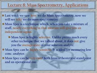
Mass Spectrometry Theory –Lecture aims. Part 1: Basic Concepts Part 2: Ion generation Part 3: Ion separation Part 4: Ion detection. Introduce basic concepts of mass spectrometry and commonly used instruments. How a Mass Spectrometer works. ION DRIFT. NEGATIVE DETECTOR.

E N D
Mass Spectrometry Theory –Lecture aims • Part 1: Basic Concepts • Part 2: Ion generation • Part 3: Ion separation • Part 4: Ion detection Introduce basic concepts of mass spectrometry and commonly used instruments
How a Mass Spectrometer works....... ION DRIFT NEGATIVE DETECTOR POSITIVE IONS _ _ _ + + + + + + _ _ _ Ions move according the Lorentz force law and Newton’s second law F = q (E + v x B) F = ma
What is Mass? Mass is given as m/z which is the mass of the ion divided by its charge Monoisotopic mass is the mass of an ion for a given empirical formula calculated using the exact mass of the most abundant isotope of each element (C=12.00000, H=1.007825 etc) Average mass is the mass of an ion for a given empirical formula calculated using the average exact mass for each element (C=12.01115, H=1.00797 etc) Nominal mass is the mass of an ion for a given empirical formula calculated using the integer mass of the most abundant isotope for each element (C=12, H=1 etc)
Isotopes and Natural Abundance • Carbon C12 (98.9%), C13 (1.1%), C14 (small) • Hydrogen H1 (99.98%), Deuterium (0.015%), Tritium (small) • Oxygen O16 (99.8%), O17 (0.04%), O18 (0.2%) • Sulphur S32 (95.0%), S33 (0.8), S34 (4.2%)
Isotope Distribution Varies with Mass Variation of Peptide Isotope Clusters Resolution with Mass
Definition of Resolution Resolution R= M1/(M2-M1) However you have to say when masses are separated….. This is usually defined as 5, 10 or 50% valley between peaks
Measuring Mass Accuracy • Measured mass • 545.4200 • Known (calculated) mass • 545.3234 • Difference • (545.4200-545.3234)/545=0.00011724 • Mass accuracy = 117 ppm 545.4200 Relative Ion Intensity m/z
Summary of Part 1: Basic Concepts • Mass is defined in three different ways • Monoisotopic, average and nominal • Isotopes define the shape of a peak • Peak shape varies with mass • Peak maximum and mass type vary with mass • Resolution defines the ability to distinguish masses • Resolution must be quoted with a qualifier • Mass accuracy can be stated in two main ways • ppm and percentage
Sample Ionisation • Derivatised small metabolites • Electron Impact (EI) • ChemicaI ionisation (CI) • Secondary ion • Peptides, proteins, RNA, DNA • Matrix assisted Laser Desorption and Ionisation (MALDI) • Electrospray ionisation (ESI) The above is also called atmospheric pressure ionisation (API). Depending on the size, it may also be called nanospray, microspray etc.
Interfacing Separation techniques with MS This involves the interfacing of a molecular separation technique to the mass spectrometer • Gas chromatography (GC) separates volatile small molecules by partitioning between the gas phase and hydrophobic solid/liquid phase • High Pressure Liquid Chromatography separates molecules by partitioning between the liquid phase and a solid phase • Capillary Zone Electrophoresis separates molecules by charge and size and can be combined with HPLC phases
Matrix Assisted Laser Desorption and Ionisation MALDI Sample: Peptide, protein, DNA… Matrix Target / Substrate:Gold / Stainless Steel / Polycarbonate / Silicon…
Electrospray Ionisation - ESI E solvent gaseous solvated ions nebulizing, drying gas needle oxidation reduction solvent 4 kV
Electrospray Ionisation and Charge [M+2H]2 + 674.7 100 Substance P % [M+H] + 685.7 693.6 666.1 600.4 1347.7 462.8 0 Da/e 300 400 500 600 700 800 900 1000 1100 1200 1300
Determining Charge State 100 95 90 524.3 85 80 75 70 65 60 55 Relative Abundance 50 45 40 35 30 525.3 25 20 15 10 526.2 5 0 520 521 522 523 524 525 526 527 528 529 Single Charge State Delta = 1.0 amu Delta = 1.0 amu Delta = 1.0 amu m/z
Determining Charge State 100 95 262.6 90 85 80 75 70 65 60 55 Relative Abundance 50 45 40 35 30 25 263.1 20 15 10 263.6 5 0 258 259 260 261 262 263 264 265 266 267 Double Charge State Delta = 0.5 amu Delta= 0.5 amu Delta = 0.5 amu m/z
Electrospray Ionisation Principles m/z = (MW + nH+) n • M2+ M3+ M+ M4+ M5+ (X+2)/2 (X+1)/1 (X+4)/4 (X+5)/5 (X+3)/3 Mass/Charge Relative Ion Intensity
Deconvoluting ESI Spectra Calc. Mass 12,055.3 Meas. Mass 12,094.4 Difference 39 (one calcium ion)
Summary of Part 2: Ion generation • Molecules must be charged • Molecules must be in the gas phase • Sample ionisation should be delicate • EI and CI are hard methods causing some fragmentation • MALDI is soft and generates mostly singly charged ions • ESI is soft and generates multiply charged ions • Ion spacing defines the charge state • Charged ion series can be deconvoluted
Ion Separation • Magnetic Sector • Quadrupoles, single and triple (SSQ, TSQ) • Ion traps (QIT) • Time-of-Flight (TOF) • Fourier Transform (FTICR-MS) • Hybrids (Q-TOF, Q-trap, TOF-TOF, trap-ICR, …)
Principle of Sector Separation Ion trajectories in a magnetic sector field Trajectories of positive ions in an electrostatic analyzer Schematic representation of a double-focusing mass spectrometer Lines marked A and B are lines of focus for direction and energy, respectively.
Quadrupole Separation Overview • The quadrupole consists of 4 parallel metal rods, or electrodes • opposite electrodes have potentials of the same sign • one set of opposite electrodes has applied potential of +[U+Vcos(ωt)] • other set has potential of -[U+Vcosωt] • U= DC voltage, V=AC voltage, ω= angular velocity of alternating voltage
Quadrupole Theory : 3 Dimensions Top and bottom electrodes Left and right ones not shown Z/Y Plane Z/X Plane Combining both planes
Quadrupole Theory: Time the fourth dimension Scanning a spectrum with paired U and V potentials
Quadruple and Trap Comparison Schematic representation of a quadrupole mass filter and an ion trap, where fo is the potential applied to opposite pairs of rods or and caps
Time of Flight Analyser (TOF): Maths + + + + • Singly charged ions generated by laser pulse • Mass proportional to final velocity hence to time of flight
Reflectron Time of Flight (ReTOF) Ion trajectories in a reflectron time-of-flight mass spectrometer, where E is the ion energy and dE the difference in ion energy of two ions
Summary of Part 3: Ion separation • There are four main types of mass spectrometers • Sector instruments (magnetic/electric) • Quadrupole (radiofrequency/voltage) • Ion traps, linear and 3D (radiofrequency/voltage) • Time of flight (voltage pulse)
Ion Detection • Point Detectors (Electron cascade) • Array Detectors (Multichannel plate) • Ion Cyclotron Resonance Cell detector
An Array Detector Bands of ions of different m/z values and separated in time travel in a broad ion beam from left to right. The ions hit the microchannel assembly and produce ion showers which are detected at the collector plate which joins them
Fourier Transform Ion Cyclotron Resonance FTMS Data Actively Shielded 7 Tesla Magnet 60 m3/hr 200L/sec 220L/sec 210L/sec 210L/sec
Trapping Ions in the ICR Cell B trapping potential
Exciting the trapped ions to a larger orbit from excite amplifier Ions circulate at their cyclotron frequencies
From “time domain” to mass spectrum Frequencies are converted into masses. m/z = k * B / f Result: A mass spectrum amplitude m/z