Download
1 / 31
310 likes | 585 Views
Practical Of Genetics. Lab. 3 Gel Electrophoresis. Objective :. To learn how to prepare agarose Gel Electrophoresis. Background. Gel electrophoresis is a widely used technique for the analysis of nucleic acids and proteins.

E N D
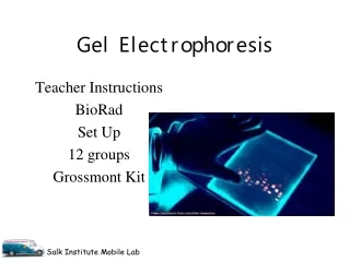
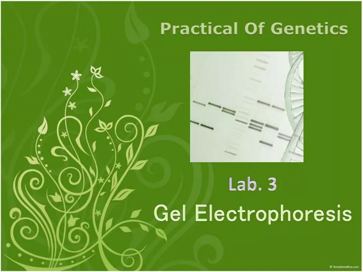
Practical Of Genetics Lab. 3Gel Electrophoresis
Objective : • To learn how to prepare agarose Gel Electrophoresis.
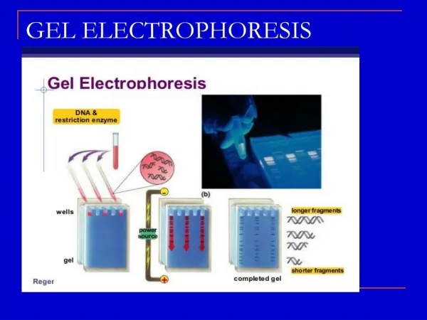
Background • Gel electrophoresis is a widely used technique for the analysis of nucleic acids and proteins. • Most every molecular biology research laboratory routinely uses agarose gel electrophoresis for the preparation and analysis of DNA.
Is a polysaccharide obtained from agar and consisting of a linear polymer (repeating units) of D-galactose and 3,6-anhydro L-galactose. • Commercially, agarose is extracted from seaweed and purified for use in electrophoresis.
The movement of molecules through an agarose gel is dependent on • The size of molecules. Small, negatively charged molecules migrate faster through agarose gels than large negatively charged molecules. • The charge of molecules. DNA, RNA, and proteins migrate toward the anode (positive electrode) when an electric field is applied across the gel.
The pore sizes present in the agarosegel. • Decreasing pore sizes increases the separation of small and large molecules during electrophoresis. • Pore size can be decreased by increasing the percentage of agarose in the gel. For example, the pore sizes are smaller in a 3% agarose gel than in a 1% agarose gel.
4) The electrophoresis buffer. • Two important parameters of the buffer are its composition and its ionic strength. • The electrical conductance of the gel is dependent on the presence of ions. Therefore, without the presence of the buffer, the current running through the gel would be very small and molecules would migrate either very, very slowly or not at all.
Conversely, a buffer with too high an ionic strength produces a very high electrical conductance and significant amounts of heat. • The heat that is produced by passing the electrical current through the gel can be hot enough to denature the DNA so that it runs through the gel as single strands instead of double strands or the heat may even melt the gel.
Is the most commonly used nucleic acid stain agarose gel electrophoresis. • Ethidium bromide intercalates double-stranded DNA and RNA. • The fluorescence of EtBr increases 21-fold upon binding to double-stranded RNA and 25-fold on binding double-stranded DNA.
Agarose • TAE Buffer • 6X Sample Loading Buffer • Ethidium Bromide (10 mg/ml) • DNA ladder standard • Electrophoresis chamber • Power supply • Gel casting tray and combs • UV light source • Gloves and goggles
CAUTION!!! This lab contains two mutagens – Ethidium Bromide (a fluorescent dye used for staining nucleic acids ) and UV light. Care should be taken when using either of these mutagens. Gloves should be worn at all times. Care should be taken never to touch gloves to notebooks, pens, benches and other surfaces. When viewing the gel with UV light, unshielded eyes should never be exposed to the UV light source. Exposed skin should also not be exposed to the UV light.
Agarose Gel Electrophoresis Protocol: • Electrophoresis buffer: usually Tris-acetate-EDTA (TAE) or Tris-borate-EDTA (TBE).
50x TAE Buffer Recipe: • Mix the following solutes and adjust to 1L by H2O. pH 8 • Store this stock solution at room temperature and dilute on your using.
10X TBE Buffer Recipe • Mix the followings and adjust the volume to 1L. Store at room temperature and dilute on your using.
6X Sample Loading Buffer • Loading buffer, which contains something dense (e.g. glycerol, sucrose) to allow the DNA sample to "fall" into the sample wells, and one or two tracking dyes (Bromophenol Blue, xylenecyanol, Orange G )which migrate in the gel and allow visual monitoring or how far the electrophoresis has proceeded. The bromophenol blue front runs at about the same position in the gel as 300 bpdsDNA and the xylenecyanol front runs at about the same position in the gel as 4,000bp dsDNA.
Measure 1g Agarose powder and add it to a 500 ml flask • Add 100 ml TAE Buffer 1X ( or TBE buffer ) to the flask. (the total gel volume well vary depending on the size of the casting tray) • Melt the agarose in a microwave until the solution becomes clear (do not let the solution boil for long periods as it may boil out of the flask). • Let the solution cool to about 50-55°C. • Add 4µl of Ethidium Bromide to the agarose solution and mix gently.
6) Seal the ends of the casting tray with two layers of tape. 7) Place the combs in the gel casting tray. 8) Pour the melted agarose solution into the casting tray and let cool until it is solid. 9) Carefully pull out the combs and remove the tape. 10) Place the gel in the electrophoresis chamber. 11) Add enough TAE buffer so that there is about 2-3 mm of buffer over the gel. 12) For example carefully pipette 10 l of each DNA sample with Loading Buffer mixture into separate wells in the gel.