Download
1 / 44
460 likes | 635 Views
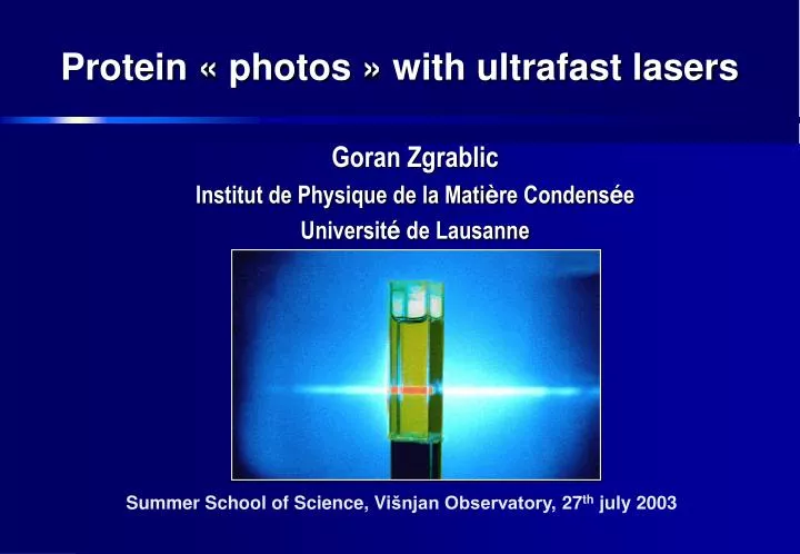
Protein « photos » with ultrafast lasers. Goran Zgrablic Institut de Physique de la Mati è re Condens é e Universit é de Lausanne. Summer School of Science, Vi šnjan Observatory , 27 th july 2003. Proteins: Nano-machines of living cell.

E N D
Protein « photos » with ultrafast lasers Goran Zgrablic Institut de Physique de la Matière Condensée Université de Lausanne Summer School of Science, Višnjan Observatory, 27th july 2003
Proteins: Nano-machines of living cell Long organic molecules which interact in biological reactions
3D Protein STRUCTURE: Methods: -X-ray diffraction -NMR (Nuclear Magnetic Resonance) -electron microscopy Protein folding still an unresolved problem “If you want to understand function, study structure” (Francis Crick) …but to know structure is not enough!
Protein DYNAMICS: the importance of the motion Time window 0.2 ns Forces: weak – comparable to thermal motions Movements: 1 Å (0.1 nm) • Molecular interactions: • Hydrophobic • Van der Waals • Electrostatic • Hydrogen bonds Aquaporine in the lipid membrane of the cell
Water and protein: a perfect couple Time window 2 ns Interactions with environment: Water is essential to biological activity of proteins Aquaporine A protein which selectively passes the water molecules into the cell (red blood cells, kidney, lung, brain, eye) Molecular dynamics simulations by B.L. de Groot and H. Grubmüller: Science 294, 2353-2357 (2001))
“If you want to understand function, study structure” (Francis Crick) Function= sequence of events over time, characterised by structural modifications “If you want to understand function, study time-dependent structures” Time resolution Spatial resolution
Conclusion:Biomolecular structure and dynamics work together to define function
Question:What do we need to make a movie of the molecules in chemical reaction?
A B AB + Fe+III [Fe+II(CN)6] [Fe+III Fe+II(CN)6]
SPECTROSCOPY UV INFRARED
The color (absorption) distinguishes reactants from product SPECTROSCOPYMeasuring absorption in time we see progress of chemical reaction
Cats are very good physicists! Time resolution 0.1 s with shutter camera
Question:What do we need to make a movie of the molecules in chemical reaction? -> LIGHT PULSE …but, how short?
The fundamental time scale in Condensed Matter, Chemistry and Biology • Speed of sound: 300 m/s-1000m/s => 0.3-1.0 Å in 100 fs • Time scale of half-oscillations: H2; we = 4155 cm-1 —> 7.6 fs I2; we = 120 cm-1 —> 270 fs 1fs = 0.00 000 000 000 000 1s = 10-15 s 1fs / 1s <=> 1s / 32 million years!
Ultrafast molecular motioninvolved in biological function 10-15 10-12 10-9 10-6 10-3 1 sec Intra-molecular motion Helix motion Protein folding P-P interaction Protein Synthesis Intermol. charge transfer Vibrations 20 … 500 fs Torsions 200 fs … 5 ps Electron transfer 20 fs … 100 psResonance energy transfer 100 fs…100 ps
Question:What do we need to make a movie of the molecules in chemical reaction? -> LIGHT PULSE of few femtoseconds
Let’s Use some light …but, somebody has to push the poor cat!
So, we need two fs pulses: t0 = 0 fs PUMPpulse – photoexcites all the molecules at the same time and starts the chemical reaction t1 = 100 fs PROBEpulse – measures the absorption change after time we want t2 = 200 fs t3 = 300 fs…
„ Femtosecond photography “ Nobel prize in Chemistry 1999: Prof. A. Zewail “ Femtochemistry “
time delay [fs] • Schémas von Selma • Pulsbreite NOPA’s
énergie distance entre les atomes
Vibration of an isolated molecule Dt = 300 fs
Femtosecond light activated processes in biologysome examples
CHROMOPHORES: Our research focuses on proteins, which can bind CHROMOPHORES (light-sensitive molecules) Molecules that react upon the exposure to light can be used as model systems for the study of these ultrafast processes
Photosensory proteins Vision Photo-taxis Plant growth Phytochrome - induction of flowering, chloroplast development, leaf senescence and leaf abscission. Understand molecular physics behind the function
all-trans 11-cis From L. Stryer, Biochemistry Rhodopsin and the retinal molecule Retinal chromophore Cis-trans isomerisation Nobel 1961: G. Wald, R.Granit, H.K. Hartline
all-trans 11-cis 200 fs Femtoseconds and proteins ?
The protein environment controls at which bond the chromophore will turn Light can stretch electron cloud when we excite chromophore Protein = has some charges and they can move around Response to photo-induced charge transfer on chromophore
+ - Amino acid measures the induced electric field... …by changing its color from blue to green
Environment (water) can make a chemical reaction possible, or make it faster
Dynamic Stokes shift – solvent responseProtonated Schiff base in MeOH
Spectrum changes in time -> water is turning around the molecule Methanol
Non-radiative energy transfer (Förster) Electron transfer in reaction center periplasm hn cytoplasm Photosynthesis From http://www.ks.uiuc.edu/Overview/gallery/structure.shtml
M. Chergui and his group IPMC - Uni Lausanne In collaboration with M. Sheves (Weizmann Institute) E. Landau (U Texas, Galveston) J. Heberle & G. Büldt (FZ Jülich) Swiss National Science Foundation Roche Research Foundation “Fondation Herbette” Lausanne Uni Lausanne