Download
1 / 69
730 likes | 1.34k Views
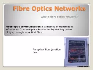
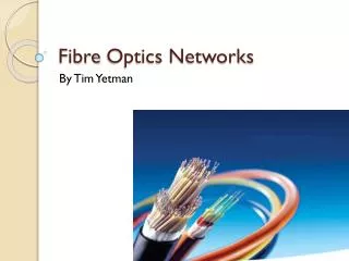
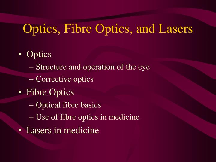
Optics, Fibre Optics, and Lasers. Optics Structure and operation of the eye Corrective optics Fibre Optics Optical fibre basics Use of fibre optics in medicine Lasers in medicine. Introduction.

E N D
Optics, Fibre Optics, and Lasers • Optics • Structure and operation of the eye • Corrective optics • Fibre Optics • Optical fibre basics • Use of fibre optics in medicine • Lasers in medicine
Introduction The term light usually refers to electromagnetic radiation in the wavelength region between about 400 and 700 nm. Although light is only a tiny part of the electromagnetic spectrum. Most of the electromagnetic radiation from the sun that reaches the Earth's surface is in this region of the spectrum. Animals have evolved light-sensitive organs which are their main source of information about their surroundings.
Introduction Vision is our most important source of information about the external world. It has been estimated that about 70% of a person's sensory input is obtained through the eye. The three components of vision are the stimulus, which is light; theoptical components of the eye, which image the light; and the nervous system, which processes and interprets the visual images
Introduction The human eye is roughly a sphere, approximately 2.4 cm in diameter. Light enters the eye through the cornea, which is a transparent section in the outer cover of the eyeball. The light is focused by the lens system of the eye into an inverted image at the photosensitive retina, which covers the back surface of the eye. Here the light produces nerve impulses that convey information to the brain.
The Eye • Structure
The Eye The focusing of the light into an image at the retina is produced by the curved surface of the cornea and by the crystalline lens inside the eye. The focusing power of the cornea is fixed. The focus of the crystalline lens, however, is alterable, allowing the eye to view objects over a wide range of distances.
The Eye In front of the lens is the iris, which controls the size of the pupil, or entrance aperture into the eye. Depending on the intensity of the light, the diameter of the aperture ranges from 2 to 8 mm. The cavity of the eye is filled with two types of fluid, both of which have a refractive index about the same as water. The front of the eye, between the lens and the cornea, is filled with a watery fluid called the aqueous humour. The space between the lens and the retina is filled with the gelatinous vitreous humour.
The Eye The focusing of the eye is controlled by the ciliary muscle, which can change the thickness and curvature of the lens. This process of focusing is called accommodation. When the ciliary muscle is relaxed, the crystalline lens is fairly flat, and the focusing power of the eye is at its minimum. Under these conditions, a parallel beam of light is focused at the retina. Because light from distant objects is nearly parallel, the relaxed eye is focused to view distant objects. In this connection, "distant" is about 6 m and beyond.
Focusing Elements • Refractive indicies within the eye Aqueous Humour 1.33 Vitreous Humour 1.33 Cornea 1.37 Crystalline Lens 1.38 (outer layers) 1.41 (inner layers)
Focusing Elements A high degree of focusing is achieved at the air-cornea interface • the crystalline lens acts more as a fine tuning element • e.g., Equivalent power of whole eye = 60 m-1 • Power of cornea = 40 m-1 • Power of lens = 20 m-1
Reduced Eye To trace accurately the path of a light ray through the eye, we must calculate the refraction at four surfaces (two at the cornea and two at the crystalline lens). It is possible to simplify this procedure with a model called the reduced eye. Here all the refraction is assumed to occur at the front surface of the cornea, which is constructed to have a diameter of 5 mm. The eye is assumed to be homogeneous, with an index of refraction of 1.333 (the same as water). The retina is located 2 cm behind the cornea.
Reduced Eye The nodal point n is the centre of corneal curvature located 5 mm behind the cornea. This model represents most closely the relaxed eye which focuses parallel light at the retina.
Reduced Eye The general lens equation is n1/u + nL/v = (nL- n1)/R1 - (nL- n2)/R2 = 1/f Here u and v are respectively the source and image distances from the lens, f is the focal length of the lens, R1 and R2 are the radii of curvature of the front and rear surfaces of the lens, n1 is the refractive index of the medium in front of the lens, n2 is the refractive index of the medium after the lens and nL is the refractive index of the lens material.
Reduced Eye For the reduced eye nL = n2 thus n1/u + nL/v = (nL- n1)/R1 = 1/f For n1 = 1 (air), nL = 1.333, R1 = 0.5cm and taking parallel incoming light (u=∞) we obtain: v = 1.333 x 5/0.333 = 20 mm Hence front focal point is 15mm in front of cornea.
Focusing Elements The viewing of closer objects requires greater focusing power. The light from nearby objects is divergent as it enters the eye; therefore, it must be focused more strongly to form an image at the retina. There is, however, a limit to the focusing power of the crystalline lens. With the maximum contraction of the ciliary muscle, a normal eye of a young adult can focus on objects about 15 cm from the eye. Closer objects appear blurred. The minimum distance of sharp focus is called the near point of the eye.
Focusing and age The focusing range of the crystalline lens decreases with age. The near point for a 10-year-old child is about 7 cm, but by the age of 40 the near point shifts to about 22 cm. After that the deterioration is rapid. At age 60, the near point is shifted to about 100 cm. This decrease in the accommodation of the eye with age is called presbyopia
Iris – Entrance Aperture of the Eye The iris is the optical aperture of the eye, and its size varies in accordance with the available light. If there is adequate light, the quality of the image is best with the smallest possible aperture. There are two main reasons for the improved image with reduced aperture. Imperfections in lenses tend to be the most pronounced around the edges. A small aperture restricts the light path to the centre of the lens and eliminates the distortions and aberrations produced by the periphery.
Iris – Entrance Aperture of the Eye A smaller aperture also improves the image quality of objects that are not located at the point on which the eye is focused. An image is in sharp focus at the retina only for objects at a specific distance from the lens system. Images of objects not at this specific plane are blurred at the retina; in other words, a point that is not in exact focus appears as a disk on the retina. The amount of blurring depends on the size of the aperture.
Iris – Entrance Aperture of the Eye A small aperture reduces the diameter of the blurred spot and allows the formation of a relatively clear image from objects that are not on the plane to which the eye is focused. The range of object distances over which a good image is formed for a given setting is called the depth of field. Clearly a small aperture increases the depth of field. It can be shown that the depth of field is inversely proportional to the diameter of the aperture.
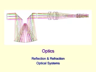
Focusing The focusing of the light into a real inverted image at the retina is produced by refraction at the cornea and at the crystalline lens. The largest part of the focusing, about two thirds, occurs at the cornea. The power of the crystalline lens is small because its index of refraction is only slightly greater than that of the surrounding fluid. The refractive power of the cornea is greatly reduced when it is in contact with water. Because the crystalline lens in the human eye cannot compensate for the diminished power of the cornea, the human eye under water is not able to form a clear image at the retina and vision is blurred.
Light Detection - The Retina The retina consists of photoreceptor cells in contact with a complex network of neurons and nerve fibres which are connected to the brain via the optic nerve. Light absorbed by the photoreceptors produces nerve impulses that travel along the neural network and then through the optic nerve into the brain. The photoreceptors are located behind the neural network, so the light must pass through this cell layer before it reaches the photoreceptors.
Fovea Retina Optic Nerve Blind Spot Light Detection - The Retina • Layer of light sensitive cells on inner surface of the eye
Light Detection - The Retina There are two types of photoreceptor cells in the retina: cones and rods. The cones are responsible for sharp colour vision in daylight. The rods provide vision in dim light. Near the centre of the retina is a small depression about 0.3 mm in diameter which is called the fovea. It consists entirely of cones packed closely together. Each cone is about 2μmin diameter. Most detailed vision is obtained on the part of the image that is projected on the fovea. When the eye scans a scene, it projects the region of greatest interest onto the fovea.
Light Detection - The Retina The region around the fovea contains both cones and rods. The structure of the retina becomes more coarse away from the fovea. The proportion of cones decreases until, near the edge, the retina is composed entirely of rods. In the fovea, each cone has its own path to the optic nerve. This allows the perception of details in the image projected on the fovea. Away from the fovea, a number of receptors are attached to the same nerve path. Hence the resolution decreases, but the sensitivity to light and movement increases.
Light Detection - The Retina With the structure of the retina in mind, let us examine how we view a scene from a distance of about 2 m. From this distance, at anyone instant, we can see most distinctly an object only about 4 cm in diameter. An object of this size is projected into an image about the size of the fovea. Objects about 20 cm in diameter are seen clearly but not with complete sharpness. The periphery of large objects appears progressively less distinct. Thus, for example, if we focus on a person's face 2 m away, we can see clearly the facial details, but we can pick out most clearly only a subsection about the size of the mouth. At the same time, we are aware of the person’s arms and legs, but we cannot detect, for example, details about the person’s shoes.
Receptors • Sensitivity:
Receptors • Dark Adaptation
Summary of Properties of Cones • “Cones” • Colour receptors (three types red, blue & green) • Respond in high illumination (daylight) • About 6.5 million per eye, concentrated at the fovea (i.e., high resolution in this region) • In the fovea, each cone connects to one nerve fibre. Elsewhere, several to one fibre • Overall peak response at ~ 550 nm
Summary of Properties of Rods • “Rods” • Respond to intensity only (monochrome) • Respond to low illumination (night vision) • About 120 million per eye, their highest concentration is at about 20º from the fovea • Hundreds of rods connect to each nerve fibre, hence low resolution • Peak response at 510 nm
Resolution of the Eye So far in our discussion of image formation we have used geometric optics, which neglects the diffraction of light. Geometric optics assumes that light from a point source is focused into a point image. This is not the case. When light passes through an aperture such as the iris, diffraction occurs, and the wave spreads around the edges of the aperture. As a result, light is not focused into a sharp point but into a diffraction pattern consisting of a disk surrounded by rings of diminishing intensity.
Resolution of the Eye If light originates from two point sources that are close together, their image diffraction disks may overlap, making it impossible to distinguish the two points. An optical system can resolve two points if their corresponding diffraction patterns are distinguishable. This criterion alone predicts that two points are resolvable if the angular separation between the lines joining the points to the centre of the lens is equal to or greater than a critical value given by sinθ = 1.22λ/d where λ is the wavelength of light and d the diameter of the aperture. For an iris diameter of 0.5 cm and green light (500nm), θ = 1.22x10⁻⁴ radians.
Resolution of the Eye Experiments have shown that the eye does not perform this well. Most people cannot resolve two points with an angular separation of less than 5 x10⁻⁴ radians. Clearly there are other factors that limit the resolution of the eye. Imperfections in the lens system of the eye certainly impede the resolution. But perhaps even more important are the limitations imposed by the structure of the retina.
Resolution of the Eye The cones in the closely packed fovea are about 2 μm in diameter. To resolve two points, the light from each point must be focused on a differentcone and the excited cones must be separated from each other by at least one cone that is not excited. Thus at the retina, the images of two resolved points are separated by at least 4 μm. A single unexcited cone between points of excitation implies an angular resolution of about 3 x 10⁻⁴ radians (using nodal point 15mm from retina). Some people with acute vision do resolve points with this separation, but most people do not. We can explain the limits of resolution demonstrated by most normal eyes if we assume that, to perceive distinct point images, there must be three unexcited cones between the areas of excitation. The angular resolution is then, as observed, 5 x 10⁻⁴ radians.
Resolution of the Eye Let us now calculate the size of the smallest detail that the unaided eye can resolve. To observe the smallest detail, the object must be brought to the closest point on which the eye can focus. Assuming that this distance is 20 cm from the eye, the angle subtended by two points separated by a distance x is: tan-1 (θ/2) = (x/2)/20. If θ is very small, this becomes θ = x/20. Because the smallest resolvable angle is 5 x 10⁻⁴ radians the smallest resolvable detail x is 0.1 mm (5 x 10-4 x 20). Using the same approach facial features such as the whites of the eye are resolvable from as far as 20m.
Sensitivity of the Eye The sensation of vision occurs when light is absorbed by the photosensitive rods and cones. At low levels of light, the main photoreceptors are the rods. Light produces chemical changes in the photoreceptors which reduce their sensitivity. For maximum sensitivity the eye must be kept in the dark (dark adapted) for about 30 minutes to restore the composition of the photoreceptors.
Sensitivity of the Eye Under optimum conditions, the eye is a very sensitive detector of light. The human eye, for example, responds to light from a candle as far away as 20 km. At the threshold of vision, the light intensity is so small that we must describe it in terms of photons. Experiments indicate that an individual photoreceptor (rod) is sensitive to 1 quantum of light. This, however, does not mean that the eye can see a single photon incident on the cornea. At such low levels of light, the process of vision is statistical.
Sensitivity of the Eye In fact, measurements show that about 60 quanta must arrive at the cornea for the eye to perceive a flash. Approximately half the light is absorbed or reflected by the ocular medium. The 30 or so photons reaching the retina are spread over an area containing about 500 rods. It is estimated that only 5 of these photons are actually absorbed by the rods. It seems, therefore, that at least 5 photoreceptors must be stimulated to perceive light.
Sensitivity of the Eye The energy in a single photon is very small. For green light at 500 nm, it is (using E=hc/λ) 4 x 10-19 Joules This amount of energy, however, is sufficient to initiate a chemical change in a single molecule which then triggers the sequence of events that leads to the generation of the nerve impulse
Vision Vision cannot be explained entirely by the physical optics of the eye. There are many more photoreceptors in the retina than fibres in the optic nerve. It is, therefore, evident that the image projected on the retina is not simply transmitted point by point to the brain. A considerable amount of signal processing occurs in the neural network of the retina before the signals are transmitted to the brain. The neural network "decides" which aspects of the image are most important and stresses the transmission of those features
Vision It has been shown that movement of the image is necessary for human vision. In the process of viewing an object, the eye executes small rapid movements, 30 to 70 per second, which alter slightly the position of the image on the retina. Under experimental conditions, it is possible to counteract the movement of the eye and stabilize the position of the retinal image. It has been found that, under these conditions, the image perceived by the person gradually fades
Limits of Detection • Lower limit of illumination • about 30 photons spread over about 500 rods • Resolution • Diffraction effects and structure of retina limit resolution to about 8mm under optimal conditions • Blind Spot • Caused by region where nerve fibres enter the optic nerve - “edited” out by the brain
Defects in Vision of the Eye There are four common defects in vision associated with the focusing system of the eye: myopia(nearsightedness), hyperopia(farsightedness), astigmatismand presbyopia (loss of accommodation) The first two of these defects are best explained by examining the imaging of parallel light by the eye.
Defects in Vision of the Eye The relaxed normal so called perfect or emmetropic eye focuses parallel light onto the retina.
Defects in Vision of the Eye In the myopic eye the lens system focuses the parallel light in front of the retina (a). This misfocusing is usually caused by an elongated eyeball or an excessive curvature of the cornea. Correction with concave lens (b).
Defects in Vision of the Eye In hyperopia parallel light is focused behind the retina (a). The problem here is caused by an eyeball that is shorter than normal or by the inadequate focusing power of the eye. Correction using a convex lens (b)
Defects in Vision of the Eye The hyperopic eye can accommodate objects at infinity, but its near point is farther away than is normal. These two defects can be summarized as follows: The myopic eye converges light too much, and the hyperopic eye not enough.