Download

1 / 16
160 likes | 164 Views
Blood culture is a laboratory test that involves injecting blood into bottles with media to determine the presence of microorganisms in the patient's blood. Learn about the purpose, pathogens that can be isolated, and the process of blood collection and examination.

E N D
Blood Culture GROUP MEMBERS: AimenNiaz RafiaHafeez AdeenaShafique ZujajatulMisbah SammiaRehman SyedaFatma H. Bukhari PRESENTED BY: AdeenaShaique SammiaRehman SyedaFatma H. Bukhari
WHAT IS BLOOD CULTURE A blood culture is the laboratory test in which blood is injected into bottles with media to determine the presence of microorganism invaded in blood of patient PURPOSE OF BLOOD CULTURE
BACTERAEMIA VS. SEPTICAEMIA • The presence of bacteria in the blood is called Bacteraemia • Bacteraemia occurs in • Typhoid fever • Brucellosis • Leptospirosis • Endocarditis. • Severe and life-threatening form of bacteraemia is called Septicaemia • Septic shock is due to • Gram negative bacilli
PATHOGENS ISOLATED FROM BLOODCULTURES • BACTERIA • E. coli • S. epidermidis • Neisseria meningitidis • Salmonella Typhi • S. aureus • FUNGI • Candida albicans • yeasts • Histoplasmacapsulatum NOTE: Blood does not have a normal microbial flora.
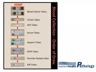
Day 1Collect blood and inoculate culture media Collection of blood: • Blood should be collected before antimicrobial treatment has started • Collect the blood as the temperature of patient begins to rise • At least two specimens (collected at different times) should be cultured. • About 20 ml of blood is taken from adults. • Blood from neonates should be collected from a peripheral vein not from the umbilical vein. • 1–2 ml of blood is enough to detect the presence of bacteria in children’s blood.
Choice of culture media • Diphasic blood culture medium • Columbia agar and broth • Commercially produced culture media NOTE: Choice of media depends on the bacterial disease that is suspected. Some bacteria grow well in a particular medium, others do not.
Aseptic blood collection and dispensingtechnique • Wash hands. • Disinfect the venepuncture site with 70% ethanol and 2% tincture of iodine. • Decontaminate blood culture bottle tops. • Using a sterile syringe, withdraw 20ml blood. • Dispense 10 ml into the culture medium. • Incubate the inoculated media as soon as possible. • Incubate upto 4 weeks when brucellosis is suspected • Incubate upto 14 days for anaerobic infections
Examining the blood culture: 1- CENTRIFUGE: a sample of EDTA anticoagulated venous blood or heparinized capillary blood and make smears of the buffy coat layers. 2- STAIN: • _ Gram smear: for Gram positive and Gram negative bacteria detection • _ Ziehl-Neelsen smear: To detect AFB (acid fast bacteria) • _ Giemsa or rapid Field’s smear: To detect borreliae, or parasites such as trypanosomes, malaria parasites, and microfilariae. 3- DRY the smears, fix with absolute methanol for 2 minutes and stain by the appropriate staining technique.
Day 2 and Onwards 3- REPORT • Diphasic culture (Columbia agar and broth) Check for microbial growth, indicated by colonies growing on the agar slope, usually beginning at the agar-broth interface. • Colonial appearances Colonies of staphylococci, S. Typhi, brucellae, and most coliforms can usually be seen easily, whereas colonies of S. pneumoniae, Neisseria species, S. pyogenes, and Y. pestis are less easily seen. • Pseudomonas and Proteus species produce a film of growth on the agar. • When growth is present: – Subculture on blood agar, chocolate agar, and MacConkey agar. – Incubate the blood agar and MacConkey agar plates aerobically and the chocolate agar plate in a carbon dioxide atmosphere (candle jar). – Examine a Gram stained smear of the colonies. • Depending on the bacteria seen, test the colonies further (e.g. for coagulase, catalase, oxidase, urease, and motility).
NOTE: When there is no growth, wash slope of diphasic culture. Reincubatecultures and subculture. • Examine toluidine blue smear (diphasic culture) • Examine thioglycollate culture • Subculture and examine microscopically • Incubate subculture anaerobically
Contamination of Blood Culture During subculturing Collection of blood • Frequent contaminants include: • Commensal staphylococci, micrococci, and diphtheroids • Contaminants from the environment • such as species of Bacillus or Acinetobacter.
Other possibilities • Occasionally in immunocompromised patients, organisms usually considered ‘contaminants’ may be pathogenic, especially fungi. • Contamination is indicated when an organism is recovered from only one bottle when it should have grown in both thioglycollate broth and the diphasicculture medium or when a mixed microbial flora is isolated.
Another use… • Severe and often fatal reactions can be caused by the transfusion of contaminated blood. • The bacteriological investigation of a transfusion reaction is as follows: