Download
1 / 17
200 likes | 403 Views
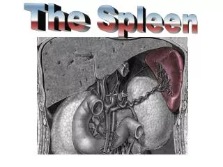
Thymus and Spleen. The Spleen: What is it good for?. Filters blood Iron Retrieval RBC reserve Immune Response* Fetal Hematopoiesis. The ‘White’ Pulp. Appears basophilic on H&E and red on silver stain Site where immune response is mounted; formation of germinal centers

E N D
The Spleen: What is it good for? • Filters blood • Iron Retrieval • RBC reserve • Immune Response* • Fetal Hematopoiesis
The ‘White’ Pulp • Appears basophilic on H&E and red on silver stain • Site where immune response is mounted; formation of germinal centers • Germinal centers with B cells and B cell derivatives push the ‘central artery’ off to the side
White Pulp Vasculature • The central artery is found in the white pulp • The central artery is surrounded by the PALS, which is T cells • Penicilli branch from the central artery into the red pulp
White Pulp Vasculature • The central artery is found in the white pulp • The central artery is surrounded by the PALS, which is T cells • Penicilli branch from the central artery into the red pulp
White Pulp Vasculature • The central artery is found in the white pulp • The central artery is surrounded by the PALS, which is T cells • Penicilli branch from the central artery into the red pulp
Red Pulp Vasculature: • Penicilli give rise to ellipsoids • Ellipsoids are capillaries ensheathed by reticular cells and macrophages; their lumens are often occluded in histo sections • Blood is filtered by macrophages through fenestrations in the sinusoids
Sinusoids See how the basal lamina is interrupted; evident with both stains
Sinusoids Lining of endothelial cells: apposed to one another, but remain separated Macrophages extend their processes into the lumen of the sinusoid (you can see the remains of RBCs in macrophages)
The Red Pulp • Appears Red on H&E • Composed of sinusoids and Cords of Billroth • The cords are the parenchyma of the red pulp; they are composed of reticular tissue w/ macrophages, red blood cells, and lymphocytes
Silver Stain *Notice how reticular fibers are evident with silver stain and not H&E *Notice the difference in appearance with the two different stains