Download
1 / 25
470 likes | 1.34k Views
Liver & Spleen. Objectives. At the end of the lecture, the student should be able to describe the: Location, subdivisions and relations and peritoneal reflection of liver. Blood supply, nerve supply and lymphatic drainage of liver

E N D
Liver & Spleen Dr. Zeenat & Dr. Vohra
Objectives At the end of the lecture, the student should be able to describe the: • Location, subdivisions and relations and peritoneal reflection of liver. • Blood supply, nerve supply and lymphatic drainage of liver • Location, subdivisions and relations and peritoneal reflection of spleen. • Blood supply, nerve supply and lymphatic drainage of liver.
Liver • The largest gland in the body • Weighs approximately 1500 g (approximately 2.5% of adult body weight). • Lies mainly in the right hypochondrium and epigastrium and extends into the left hypochondrium. • Protected by the thoracic cage and diaphragm, lies deep to ribs 7-11 on the right side and crosses the midline toward the left nipple. • Moves with the diaphragm • Located more inferiorly when one is erect because of gravity.
Relations of Liver • Anterior: Diaphragm, Right and left costal margins, right and left pleura and lower margins of both lungs, xiphoid process, and anterior abdominal wall in the subcostal angle • Posterior: Diaphragm, right kidney, hepatic flexure of the colon, duodenum, gallbladder, inferior vena cava, and esophagus and fundus of the stomach
Peritoneal Reflection Superior layer of coronary ligament Diaphragm Peritoneum Inferior layer of coronary ligament Anterior abdominal wall Posterior abdominal wall Other bare areas include portahepatis; fossa for gall bladder & grooves for IVC • The liver is completely surrounded by a fibrous capsule and covered by peritoneum (except the bare areas). • The main bare area of the liver is an area on the diaphragmatic surface where there is no intervening peritoneum between the liver and the diaphragm • Boundaries of Bare area: • Anterior: superior layer of coronary ligament • Posterior: inferior layer of coronary ligament • Laterally: right and left triangular ligaments
Surfaces of Liver • The liver has two surfaces: • A convex diaphragmatic surface • A relatively flat or even concave visceral surface (posteroinferior)
Diaphragmatic Surface • The convex upper surface is smooth and molded to the undersurface of the domes of the diaphragm which separates it from the pleurae, lungs, pericardium, and heart . • Covered with visceral peritoneum, except posteriorly in the bare area of the liver, where it lies in direct contact with the diaphragm.
Visceral Surface • It is the posteroinferiorsurface, related to abdominal viscera. • It is covered with peritoneum, except at the fossa for the gallbladder, groove for IVC and the portahepatis • It bears multiple fissures and impressions from contact with other organs.
Fissures • Two sagittally oriented fissures, linked centrally by the transverse portahepatis, form the letter H on the visceral surface. • The left fissureis the continuous groove formed: • Anteriorly by the fissure for the round ligament • Posteriorly by the fissure for the ligamentumvenosum. • The right fissureis the continuous groove formed: • Anteriorlyby the fossa for the gallbladder • Posteriorly by the groove for the inferior vena cava. Inferior vena cava Ligamentumvenosum Portahepatis Round ligament Gall bladder
Relations of Visceral Surface of the Liver 1 2 6 3 4 5 The visceral surface is related to the: stomach Esophagus lesser omentum gallbladder right colic flexure right kidney and right suprarenal gland
PortaHepatis (Hilum of the Liver) • Structures passing through the portahepatis include: • Right and left hepatic ducts • Right and left branches of the hepatic artery • Right and left branches of the portal vein • Sympathetic and parasympathetic nerve fibers • A few hepatic lymph nodes lie here; they drain the liver and gallbladder and send their efferent vessels to the celiac lymph nodes. • A transverse fissure found on the posteroinferior surface and lies between the caudate and quadrate lobes. • The upper part of the free edge of the lesser omentum is attached to its margins.
Ligaments of the Liver • Falciform ligament • It is a two-layered fold of the peritoneum, ascends from the umbilicus to the liver • Its sickle-shaped free margin contains the ligamentumteres (round Ligament) of liver, the remains of the umbilical vein,which carried oxygenated from the placenta to the fetus. Ligamentumvenosum IVC PortaHepatis Round ligament of liver GB Ligamentumvenosum Itis the fibrous remnant of the fetal ductusvenosus, which shunted blood from the umbilical vein to the IVC, short-circuiting the liver.
Lobes of The Liver Caudate process Cauate lobe Left lobe Right lobe Quadrate lobe The liver is divided into a large right lobe and a small left lobe by the attachment of the falciform ligament. The right lobe is further divided into a quadrate lobe and a caudate lobe by the presence of the gallbladder, the fissure for the ligamentumteres, the inferior vena cava, and the fissure for the ligamentumvenosum. The caudate lobe is connected to the right lobe by the caudate process. The quadrate and caudate lobes are a functional part of the left lobe of the liver.
Blood Circulation through the Liver • The blood vessels conveying blood to the liver are the hepatic artery (30%) a branch of celiac trunk, and portal vein (70%). • The hepatic artery brings oxygenated blood to the liver • The portal vein brings venous blood rich in the products of digestion, which have been absorbed from the gastrointestinal tract. The venous blood is drained by right & left hepatic veins which drain into the inferior vena cava
At or close to the portahepatis, the hepatic artery and portal vein terminate by dividing into right and left primary branches which supply the right and left parts of liver, respectively. • Within the liver, the primary branches divide to give secondary and tertiary to supply the hepatic segments independently • The hepatic veins, are intersegmental in their distribution and function, draining parts of adjacent segments. • The attachment of these veins to the IVC helps hold the liver in position.(The peritoneal ligaments and the tone of the abdominal muscles play a minor role in the support of liver).
Lymph Drainage • The liver produces a large amount of lymph—about one third to one half of all body lymph. • The lymph vessels leave the liver and enter several lymph nodes in the portahepatis. • The efferent vessels pass to the celiac nodes. • A few vessels pass from the bare area of the liver through the diaphragm to the posterior mediastinal lymph nodes. • Nerve Supply Sympathetic and parasympathetic nerves from the celiac plexus. The anterior vagal trunk gives rise to a large hepatic branch, which passes directly to the liver.
Portal-Systemic (Portacaval) Anastomoses • It is a specific type of anastomosis that occurs between the veins of portal circulation and those of systemic circulation • In portal hypertion, these anastomosis open and form venous dilatations called varices. • Sites: • Esophagus • Anal canal • Paraumbilical region • Retroperitoneal • Intrahepatic (Patent ductusvenosus)
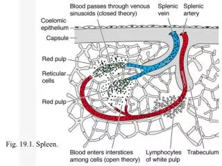
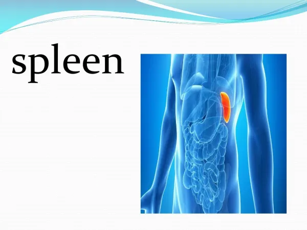
Spleen • Largest single mass of lymphoid tissue • Located in the left hypochondrium, deep to 9, 10 & 11 ribs • Long axis lies along the shaft of the 10th rib and separated from them by the diaphragm and the costodiaphragmatic recess • Ovoid in shape with notched anterior border • Lower pole extends forward as far as the midaxillary line • Normal size spleen can not be palpated on clinical examination
Surfaces: • Diaphragmatic surface:is convexly curved to fit the concavity of the diaphragm and curved bodies of the adjacent ribs • Visceral surface: related to viscera Borders: The anterior and superior borders are sharp. Anterior border is notched The posterior (medial) and inferior bordersare rounded
Peritoneal Reflections/Ligaments • Spleen is completely surrounded by peritoneum which passes from it at the hilus as: • Gastrosplenic ligament to the greater curvature of stomach (carrying the short gastric and left gastroepiploic vessels) • Lienorenal ligament to the left kidney (carrying the splenic vessels and the tail of pancreas)
Relations • Anteriorly: Stomach, tail of pancreas, left colic flexure & left kidney • Posteriorly: Diaphragm, that separates it from the left pleura (left costo-diaphragmatic recess), left lung & 9, 10 & 11 ribs • Inferiorly: Left colic flexure. • Medially: Left kidney.
Arterial Supply Splenic artery • Largest branch of the celiac artery • Runs a tortuous course along the upper border of the pancreas • Passes within the lienorenal ligament • Divides into 4-5 branches, which enter the spleen at the hilus • The lack of anastomosis of these arterial vessels within the spleen results in the formation of vascular segments of the spleen with relatively avascular planes between them, enabling subtotal splenectomy
Venous Drainage Splenic vein • Leaves the hilus • Runs behind the tail & body of the pancreas • Reaches behind the neck of pancreas, where it joins the superior mesenteric vein to form the portal vein • Tributaries: • Short gastric vein • left gastroepiploic vein • Pancreatic veins • Inferior mesenteric vein
Lymph Drainage • Nerve Supply • Lymphatics emerge from the hilus and drain into several nodes lying at the hilum • Efferents from the hilar nodes pass along the course of splenic artery, and drain into the celiac lymph nodes Derived from the celiac plexus Are distributed mainly along branches of the splenic artery, and are vasomotor in function.