Download
1 / 79
800 likes | 987 Views
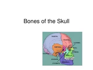
NEUROLOGY REVIEW The bare bones of the thing. Cerebrovascular Disease Neurodegenerative Conditions Epilepsy Demyelinating Disease Neuropathies Headaches Neuromuscular Conditions. Cerebrovascular Disease.

E N D
NEUROLOGY REVIEWThe bare bones of the thing Cerebrovascular Disease Neurodegenerative Conditions Epilepsy Demyelinating Disease Neuropathies Headaches Neuromuscular Conditions
Cerebrovascular Disease • Risk factors: Age, Family Hx, HTN, Cholesterol, Diabetes, Heart Disease (esp Afib, cardiomyopathy, atrial thrombus, PFO), tobacco use, excessive etoh use, hypercoagulable states (cardiolipin syndrome, CA, anti-thrombin 3 deficiency, prot C and prot S deficiency)
Ischemic strokes of sudden onset or stuttering in onset Deficits worse at onset with gradual improvement Most common type(85%) Hemorrhagic strokes of sudden onset Deficits worsen with time Headache is common Additional etiologies: AVM, aneurysm, amyloid angiopathy Ischemic vs Hemorrhagic
Thrombotic vs Embolic • Clinically, impossible to differentiate • Most strokes are thrombotic (?80%) • If potential source is identified (Carotid stenosis, Afib, atrial thrombis, cardiomyopathy, endocarditis, PFO, aortic arch plaque, dissection) then assume embolism. Otherwise, assume intracranial thrombosis.
Left Hemisphere • Right hemiparesis, hemisensory deficit • Aphasia if cortical location • Hyper-reflexia on right, Babinski on right • Initially flaccid tone with eventual spasticity on right
Right Hemisphere • Often silent or less pronounced symptoms due to the dominance of the left hemisphere • Left hemiparesis, hemisensory deficit • Anosognosia • Hyper-reflexia on left with left Babinski • Initially flaccid with eventual spasticity on left
Occipital infarcts • Left or right hemi-visual field loss • Headache common • Patient often misinterprets that one eye doesn’t work. The examiner needs to discern the truth by checking visual fields
Cerebellar infarcts • Objectively: Ataxia, dysdiadachokinesis, tremor, dysarthria, nystagmus • Subjectively: Dizziness, nausea, clumsiness • Higher risk of hemorrhagic transformation compared to other brain areas
Brainstem Infarcts • Cranial nerve signs: diplopia, facial numbness, facial weakness, ptosis, pupil irregularities, dysphagia, asymmetric palate or tongue, • Crossed motor or sensory deficits, or even biparesis or quadraparesis
Transient Ischemic Attacks • Three minutes or less. This is a meaningless distinction. The event is still a stroke. • Most significant is Amaurosis Fugax. “Fleeting Blindness” may represent embolic event from carotid stenosis • Any TIA is a harbinger of another stroke in the near future (15% chance within a month)
Workup • MRI brain is preferable. CT’s are quick • Carotid Duplex. MRA or CT angiogram if dissection is suspected • TTE or TEE • EKG/Holter • Hypercoagulation blood work if indicated
Emergent Treatment • Anti-platelet medication at onset (except, of course, in hemorrhage) • Anti-coagulation if embolism from Afib, atrial thrombus, or cardiomyopathy, or dissection is determined • tPA if available within three hours of onset.
Prevention • Anti-platelet medication in almost all cases • Anti-coagulation only in Afib, cardiomyopathy, atrial thrombus, “shaggy aorta syndrome” • Carotid endarterectomy if ipsilateral stenosis greater than 70% • PFO closure • Treatment of underlying risk factors
Neurodegenerative Conditions • Alzheimer’s Disease • Parkinson’s Disease • Huntington’s Disease • Progressive Supranuclear Palsy • Cerebellar Degeneration
Alzheimer’s Disease • Estimated 50% of people age 85 years • Familial if parent had disease in early to mid 50’s • Risk factors: loose association to past head injuries, inverse association to IQ and education level
AD Pathology • Neurofibrilary tangles, neuritic plaques • Generalized brain atrophy as disease progresses, but especially in temporal lobes • Reduction in acetylcholine system
AD symptoms/signs • Progressive “short term” memory deficits, i.e. inability to form new memories. • Progressive aphasia • Personality change • Progressive deficits in ADL’s - Apraxia
AD Workup • Brain imaging • B12 level, TSH • Mini Mental Status exam • Clock Drawing
AD continued • The leading cause of dementia in the USA is Alzheimers. • The second leading cause is multiple strokes • The third cause is a combination of the two above.
AD Treatment • Acetylcholine esterase inhibitors - Aricept, Exelon, Razadyne • Glutamate inhibitor - Namenda • These drugs only slow the progression of the disease, they do not lessen the deficits • In this disease, you treat the family perhaps more than the patient
Parkinson’s Disease • Very common - estimated one in one hundred between ages 60 and 70 years • Tends to be sporadic • Rate of disease progression is very variable
PD Pathology • Progressive degeneration of pigmented dopamine producing cells in the substantia nigra, i.e. nuclei in the mid-brain • Degeneration begins at birth. • Symptoms appear when degeneration reaches 80%
PD Symptoms/Signs • Tremor - Resting. Often begins unilaterally in extremities. Can involve the jaw and tongue • Rigidity - increased tone in the extremity muscles. Cogwheeling if superimposed tremor. Stooped posture • Akinesia (bradykinesia) • Postural Instability
PD Symptom/Signs continued • Drooling, constipation, micrographia, low volume stuttering speech, dementia, reduced facial expression, reduced eyeblink rate
PD Treatment • Neuroprotective agents: Vitamin E, CoEnzyme Q10 • Dopamine agonists: Requip, Mirapex, Permax. More useful early on when there are more dopaminergic cells still functioning • Anti-cholinergics especially for tremor • Dopamine replacement: Sinemet (carbidopa/levadopa). Gold standard treatment (high risk of adverse effects at five years) - Less is Best • Deep Brain Stimulation
Huntington’s Disease • Autosomal Dominant disorder. • Easily diagnosed via blood test that detects the genetic defect, i.e. CAG repeat sequence - the longer the sequence, the earlier onset and more severe the symptoms • Symptoms usually manifest in young adulthood
HD Clinical triad • Chorea - involuntary, writhing or quick movments of face, limbs, trunk • Psychiatric disease - usually depression and can occur before other deficits • Dementia - usually occurs later
HD Treatment • Is a progressive disease. Life expectancy average 19 years • No known treatment, except to treat the depression when needed or to reduce the chorea with dopamine blockers. Dementia is resistant to treatment • Again, treat the family
Progressive Supranuclear Palsy • Usual onset 6th to 7th decade • Sporadic • Pathology - degeneration of brain stem nuclei in midbrain “and above” to the basal ganglia • Often mistaken for Parkinson’s Disease because of the bradykinetic appearance of the patient
PSP Continued • Hallmark Sign: impaired voluntary vertical eye movements • Also, axial more than appendicular rigidity, grimacing, drooling, dysarthria • Prominent early symptom: Falling especially going down steps (due to impaired vertical eye movements)
PSP continued • Treatment - none • This is another disease where the treatment is directed more to the family than the patient
Cerebellar Degeneration • Autosomal recessive types tend to occur early adulthood • Autosomal dominant types tend to occur later life • Both are slowly progressive and similar in deficits
CD continued • Progressive ataxia, dysarthria, irregular tremor in extremities and trunk (titubation), dysphagia, nystagmus • Dementia is subtle and generally not significant
Epilepsy • A seizure is a single event due to an abnormal discharge of a group of cerebral neurons resulting in a multifarious array of behaviors and/or cognitive experiences • There are many etiologies for seizures - trauma, hypoxia, drugs, fever, etc.
Epilepsy Continued • Epilepsy is a disorder of the brain that produces recurrent spontaneous, seizures. • Two age peaks - childhood to young adult and later life (age 55+) • Younger group has 5-20%chance of resolution and is often hereditary. Older group has little to no chance of resolution and is usually sporadic.
Types of seizures • Simple Partial - motor, sensory, visual, cognitive or emotive event (depending on location of the electrical discharge) WITHOUT change in consciousness • Complex Partial - the above WITH change in consciousness • Generalized Tonic-Clonic Seizure • Absence, Myoclonic, Atonic, Tonic, Clonic seizures
Types of Epilepsy • Partial Epilepsies (location specific) result in simple partial, complex partial or secondarily generalized tonic-clonic convulsions. An abnormal electrical focus in the brain may be due to scar, tumor, congenital malformation • Primary Generalized Epilepsies result in absense, myoclonic, tonic, clonic seizures or tonic-clonic convulsions. No abnl focus is found. Rather, the brain has lower seizure threshold.
Epilepsy workup • Obtain as complete a history as possible • EEG to determine type of Epilepsy (recall EEG’s only 90+% sensitive) • Brain imaging re possibility of mass, stroke, etc. • Select urine tox screen or blood studies as indicated
Epilepsy Treatment • Medication choice depends, in part, on type of Epilepsy • Primary Generalized Epilepsies respond best to Depakote, Lamictal, Keppra and Topamax. Avoid Tegretol. • The Partial Epilepsies respond to all medications
Epilepsy treatment continued • The Generalized Epilepsies are usually more successfully treated, whereas the Partial epilepsies are more challenging. • 70% of people are 100% controlled on monotherapy. The remainder require polypharmacy and may not have 100% control • Vagus Nerve Stimulation and lesionectomy or corpus callosotomy are used in refractory cases.
Epilepsy medication side effects • Depakote - spina bifida, weight gain, tremor, hair loss • Tegretol - hyponatremia • Dilantin - gingivitis, hirsutism, osteoporosis • Topamax - renal calculi • Phenobarbitol - sedation and cognitive impairment
Epilepsy and pregnancy • Overall risk to mom and baby is greater if mom is untreated. • Birth defect rate general population 2% • Birth defect rate untreated epileptics 4% • Birth defect rate treated epileptics 6% • Supplemental folate, monitor drug levels, mom considered “high risk”
Status Epilepticus • Thirty minutes or more of continuous seizing or recurrent seizures without recovery between them. • Is an emergency because of potential neuronal damage due to high metabolic activity and hypoxia. • Rx: Benzo first followed by IV Fosphenytoin and possibly IV phenobarbitol
CNS Demyelinating Disease • Multiple Sclerosis is an auto-immune disease affecting both the brain and spinal cord in which myelin is attacked by anti-bodies. This results in destruction of the myelin and if severe can lead to axonal destruction and ultimately neuronal death. • “Sclerosis” refers to the calcified scarring seen in multiple areas of the brain and/or spinal cord at autopsy
Demyelinating Disease continued • If only the spinal cord is affected, the condition is referred to as Transverse Myelitis • Once the brain is affected , the condition is referred to as Multiple Sclerosis • It is a disease primarily of young women
MS - Course of the disease • Relapsing-Remitting 70% • Primary Progressive 10% • Secondary Progressive - about 50% of the R-R type at 15 years • Monosymptomatic - ?10% • Devic’s Disease - TM plus ON only 5%
MS Workup • MRI showing multiple (at least nine) peri-ventricular lesions typically perpendicular to ventricles, some of which enhance • Lumbar Puncture revealing oligoclonal bands, myelin basic protein, elevated CSF/serum IgG ratio • Visual Evoked Potential • Clinical criteria “dissemination in time and space.” This is still the best way to diagnose
MS Symptoms/Signs • Typical relapse is gradual onset (2d to 2w) of a neurologic deficit, plateau (2w to 2m) and gradual recovery • Deficits include motor, sensory, balance, visual (Optic Neuritis in particular), speech, cognitive problems • Fatigue, depression, dementia common and long lasting • Average 2 relapses per year
MS Treatment • Steroids useful in shortening the duration of a particular relapse and in reducing its’ severity • Immunomodulating medications useful in reducing relapse rate, reducing “lesion load” in the CNS, and ultimately in reducing long term disability
MS Treatment continued • Currently six options • Beta seron - LFT elevations, flu-like symptoms, leukopenia • Avonex - as above but less problematic • Rebif - very similar to Beta seron • These three are all Interferons and suppress B cell antibody production.Higher potency interferons are more efficacious but carry increased adverse effects.