Download
1 / 64
830 likes | 1.55k Views
Diseases of the Aorta. Seoul National University Hospital Department of Thoracic & Cardiovascular Surgery. Anatomy of Aorta. Aortic root aortic valve, sinus of Valsalva, coronary artery Ascending aorta aortic root ~ innominate artery Aortic arch proximal, distal

E N D
Diseases of the Aorta Seoul National University Hospital Department of Thoracic & Cardiovascular Surgery
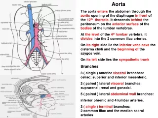
Anatomy of Aorta • Aortic root • aortic valve, sinus of Valsalva, coronary artery • Ascending aorta • aortic root ~ innominate artery • Aortic arch • proximal, distal • Descending thoracic aorta • distal to LSCA ~ 12th ICS • Thoracoabdominal aorta • descending thoracic aorta & abdominal aorta
Properties of Aorta & Major Conduit 1. Aorta Compliant vessel (Windkessel function) ; transforms pulsatile hydraulic energy into a more steady flow by elastic distension & contraction 2. Synthetic conduit Noncompliant ; must result in alteration of arterial hemodynamics & LV load (increased impedance & afterload)
Diseases of Thoracic Aorta • Aortic aneurysm • Aortic dissection • Obstructive disease of branches of the thoracic aorta • Traumatic aortic rupture
Pathophysiology of Aortic Aneurysm • Definition • localized or diffuse dilatation > 50% of normal diam. • Most common aortic disease that require surgery • Etiology • Atherosclerosis ( + underlying weakness) • Chronic aortic dissection • Annuloaortic ectasia (Marfan syndrome) • Trauma • Infection • Associated with aortic valve disease
Histopathology of Ascending Aortic Aneurysm 1. Cystic medial necrosis by pooling of mucoid material 2. Elastin fragmentation by disruption of elastin lamellae 3. Fibrosis as an increase in collagen at the expense of smooth muscle cells 4. Medionecrosis as areas with apparent loss of nuclei
Pathophysiology of Ascending AA • Marfan syndrome • Incidence • 1 / 5,000 • Annuloaortic ectasia is very common • Associated defects • Aortic regurgitation, mitral valve prolapse, dysrhythmia • Tall stature, long limbs and digits, anterior chest deformity, joint laxity, vertebral column deformity • High arched palate, lens disorder
Marfan’s Syndrome * Definition • A heritable disorder (AD) of connective tissue involving biochemical abnormality of extracellular matrix by a mutation in fibrillin gene on chromosome 15 (Fibrillin-1, 350-KD glycoprotein : integral structural component of 10-nm noncollagenous microfibrils of extracellular matrix in most tissue) • The absence of structural integrity of skeletal, ocular, & cardiovascular system • Adult patients demonstrate abnormal elastic properties manifested by decreased aortic distensibility & increased stiffness index
Marfan’s Syndrome • Clinical manifestations • Cardiovascular • Ocular • Skeletal abnormality • Cardiovascular manifestations • Progress with time • Mitral valve prolapse in 100% • Aortic root dilatation in 80% • Rarely atrial septal aneurysm
Manifestations of Marfan’s Syndrome • 1. Patterns of aortic dilatation • 1) 80% of the patients shows aortic dilation • 2) more commonly generalized form than localized form • 3) more commonly aortic regurgitation in generalized form • 2. Natural prognosis 1) Life expectancy is significantly reduced (40~50) as a consequence of aortic dilatation & its complications (aortic dissection, fatal rupture, AR, heart failure)
Marfan’s Syndrome in Children • 1. Diagnosis can be made at any age with marked variation • in clinical expression. • 2. Patients without family history (in one third of patients • of all age) have more severe manifestation probably • due to sporadic mutation. • 3. Surgery should be carried out even in asymptomatic • patients, once the diameter of the aortic root or • ascending aorta reaches 5 to 6cm as in adults. • 4. Mitral valve prolapse is as common as aortic root • dilatation and progression can cause significant • morbidity & mortality.
Patterns of Aortic Aneurysm • Locations of Aneurysm • Ascending aorta 45 % • Aortic arch 10 % • Descending thoracic aorta 35 % • Thoracoabdominal aorta 10 %
Natural History of AA • Aortic aneurysm • Incidence • 5.9 new aneurysms / 100,000 person-years • Life time probability of rupture : 75~80% • 5-yr untreated survival rate : 10~20% • Median time to rupture : 2~3 yrs Size Risk of rupture within 1yr < 5 cm 4 % 6 cm 43 % 8 cm 80 %
Clinical Presentation of AA • Symptoms & signs • Asymptomatic • Compressive symptoms • recurrent laryngeal n. or vagus n. : hoarseness • tracheobronchial tree : dyspnea • pulmonary a. : fistula, bleeding pulmonary HT & edema • esophagus : dysphagia • stomach : sensation of satiety wt. loss • Pain aneurysmal expansion • Intestinal angina, renovascular HT associated atherosclerotic obstructive disease (5% in TAAA) • Physical finding-usually unremarkable • Wide pulse pressure, diastolic murmur AR
Indications for Aortic Aneurysm • Aneurysm diameter 5cm • Aneurysm with documented enlargement • Symptomatic aneurysm ― chest pain or back pain indicating expansion ― significant aortic regurgitation
Dissecting Aortic Aneurysm • Catastrophic event • Intimal tear • False channel • in the outer half of the media • highly susceptible to rupture • Acute dissection < 2 wks from Sx onset • Chronic dissection > 2 wks from Sx onset
Pathophysiology of Aortic Dissection Malperfusion Reentry
Predisposing Factors of DA • Hypertension • Cystic medial necrosis • Marfan syndrome • AAE(annuloaortic ectasia) • Bicuspid aortic valve • Coarctation • Pregnancy • Chest trauma
Classification of Dissection • Standford • Type A • Involvement of the a-Ao ( arch or d-Ao) • regardless of site of primary intimal tear • Type B • All others without involvement of a-Ao • DeBakey I, II, III • According to the location of intimal tear
Classification of Aortic Dissection II I III B A
Natural History of DA • Annual incidence • 5~10 / million • Sex ratio • M:F = 2:1 ~ 5:1 • Acute dissection • Median time to rupture : 3 days • Mortality rate ; 50 % within 2 days 75 % within 2 wks • Chronic dissection • Median time to rupture : 1~3 Yrs • Follows patterns of non-dissecting aneurysm
Clinical Presentation of DA • Acute dissection • Excruciating pain • abrupt onset • sudden rise to peak • Chest pain • 2/3 of a-Ao dissection • Back pain • dissection distal to aortic arch • Pain may migrate as the dissection moves distally. • Various extent of peripheral & central vessel occlusion • from progression of dissection through the false lumen • Failure of diagnosis : major problem
Clinical Presentation of DA Type A Type B Frequency Pain anterior substernal posterior, midscapular, abdominal Syncope +++ rare Dyspnea + ― Blood pressure elevated 50%, low 20% elevated 80% Asymmetric pulses upper, lower extremity lower extremity 30-50% Diastolic murmur 50% 10% Pericardial effusion +++ rare Pleural effusion ± +++ Hemiparesis or plegia + ― 5-6% Paraparesis or plegia + + 2-6% Renal, intestinal infarction + + 3-5% Myocardial infarction + rare 10 %
Principle of Treatment in DA • Type A acute aortic dissection • Emergent operation • Type B acute aortic dissection • Medical Tx and observation unless life threatening • Surgical indication • Persistent pain • Aneurysmal dilatation ( 5cm) • End organ (kidney, bowel) or limb ischemia • Evidence of retrograde dissection to the a-Ao
Medical Management of DA • Initial management • Immediate ICU care • BP control & Monitoring • Central line, arterial line, urine output • Imaging studies • Daily Chest X-ray, weekly CT scan during hospitalization • Pharmacologic therapy • Vasodilator : Sodium nitroprusside • β-blocker : Esmolol (β-1 selective & short acting)
Diagnostic Studies for DA • CT & CT angiography • Aneurysm size, location, extent, intimal tear site • Other pathologies in the chest & abdomen • Follow-up study : aneurysm growth • Limitation • unreliable detection of root enlargement • Contraindication • renal insufficiency, allergy to contrast agents
Diagnostic Studies for DA • MRI • Noninvasive study • Do not require contrast medium • Better than CT at detecting aortic root dilatation • Disadvantages • cost • required time (esp, in acute dissection) • Contraindication • pacemaker, claustrophobia
Diagnostic Studies for DA • Transesophageal Echocardiography (TEE) • Accuracy in imaging intimal tear : 90% • Assessment of cardiac structure & function • Highly sensitive in aortic pathology diagnosis • aortic valve disease, aortic dilatation, dissection, thrombi, atherosclerotic disease • Intraoperative monitoring • check cardiac function, aortic valve competency, atherosclerosis in the thoracic aorta • Limitation • requires a skilled cardiologist
Diagnostic Studies for DA • Aortography • Geography of the aorta & condition of smaller vessels • Previous gold standard in dissection • double lumen, tear site, extent • Indication • renovascular HT, intermittent claudication, atherosclerotic occlusive abdominal aorta, symptoms of carotid artery occlusion • Disadvantages • invasive procedure using radiopaque dyes • Cardiac cath & coronary angiography • Evaluation of the concomitant coronary artery disease
Surgery of Type A Dissection • Principles of Surgical Tx in Acute Dissection • Resection of aortic segment containing intimal tear • Obliteration of false lumen in both end of remained aorta • Graft replacement of resected aortic segment • Techniques • Median sternotomy • Femoral-femoral bypass • Trendelenburg position • Circulatory arrest with deep hypothermia • Retrograde cerebral perfusion • Reinforcement of the intima & adventitia together (sandwich technique)
Surgery of Acute Type B Dissection • Techniques • Similar to the techniques for aneurysm • Rechanneling blood into the true lumen • Ligation of all intercostal arteries in acute dissection • Surgical indications • Persistent pain • Aneurysmal dilatation ( 5cm) • End organ(kidney, bowel) or limb ischemia • Evidence of retrograde dissection to the a-Ao
Surgical Results of DA • Acute Type A Dissection • Early mortality : 20~30 % • Main cause of death underlying end-organ injury • Major complications stroke (9%) • Major risk factors for postop. stroke • pump time, episode of severe hypotension • Acute Type B Dissection • Early mortality : 25~50 % (cf. medical treatment : 7~32 %) • Major complications : ischemic spinal cord injury
Surgical Treatment of AA Aneurysm : Aortic Root, a-Ao, Aortic Arch • Historical evolution • 1950s : Cardiopulmonary Bypass (Gibbon) • 1955 : 1st successful a-Ao repair (Cooley & DeBakey) • 1964 : 1st successful replacement of entire a-Ao (Wheat) • CPB, coronary perfusion, myocardial cooling, cold cardiac arrest • 1968 : Composite valve graft (Bentall & de Bono) • 1975 : Replacement of entire aortic arch (Griepp) • profound hypothermia & circulatory arrest
Aortic Root, Ascending Aorta, Aortic Arch • Limitation of profound hypothermia < 30 min : safe duration > 45 min : increased incidence of stroke > 65 min : increased incidence of death • Calculated safe duration of hypothermic circulatory arrest Temperature Cerebral Metabolic Rate Safe Duration of HCA (C) (% of baseline) (min) 37 100 5 30 56 ( 52 ~ 60 ) 9 ( 8 ~ 10 ) 25 37 ( 33 ~ 42 ) 14 ( 12 ~ 15 ) 20 24 ( 21 ~ 29 ) 21 ( 17 ~ 24 ) 15 16 ( 13 ~ 20 ) 31 ( 25 ~ 38 ) 10 11 ( 8 ~ 14 ) 45 ( 36 ~ 62 )
Aortic Root, Ascending Aorta, Aortic Arch • Adjuncts for brain protection • Reintroduction of antegrade cerebral perfusion (Frist, 1987) • Retrograde cerebral perfusion (Ueda, 1989)
Aortic Root - Techniques • Median sternotomy • Antegrade and/or retrograde cardioplegic perfusion • Techniques for aortic root • Wheat • Composite graft (esp, for Marfan) • Bentall • Cabrol • modified Cabrol • button • Homograft • Valve sparing procedure Choice of tube graft ; diameter of 10% smaller than the length of the free margin of the aortic leaflet
Valve-sparing Operation Resorting aortic root dimensions in an aortic valve-sparing operation when aortic annulus is normal and sinotubular junction is enlarged
Valve-sparing Operation Resorting aortic root dimensions when aortic annulus & sinotubular junction are normal, as in aortic dissection
Valve-sparing Operation Resorting aortic root dimensions when aortic annulus and sinotubular junction are enlarged, as in anuloaortic ectasia with Marfan syndrome
Valve-sparing Operation Reconstructing aortic root using a graft with the aortic valve placed within it
Aortic Root – Wheat Technique • Separate valve/graft replacement • For older patients with mild to moderate sinus dilatation
Aortic Root – Composite Valve Graft • Bentall technique • Coronary artery reattachment side-to-side anastomosis • Disadvantage bleeding d/t anastomosis tension → pseudoaneurysm (7~25%)
Aortic Root – Composite Valve Graft • Cabrol technique • Coronary artery reattachment • a small graft to the both coronary arteries • side-to-side anastomosis of the small graft & composite graft • Advantage • ↓anastomosis tension • Disadvantage • kinking at the anastomosis sites
Aortic Root – Composite Valve Graft • Modified Cabrol technique • Coronary artery reattachment • a small graft to the LCA • end-to-side anastomosis of the small graft & composite graft • button attachment of the RCA • Advantage • ↓kinking
Aortic Root– Composite valve graft • Button technique • Coronary artery reattachment • Carrel patch for both coronary a. • Direct anastomosis to the composite graft
Composite Valve Graft A; aortic valve is excised B; composite prosthetic valve conduit is attached to annulus of aortic valve
Surgery of Aortic Root • Results • Early mortality : 2~15% • Early complications : thromboembolism, bleeding • Late complications : endocarditis, thromboembolism pseudoaneurysm
Surgery of Aortic Root • Results