Download
1 / 134
1.38k likes | 1.62k Views
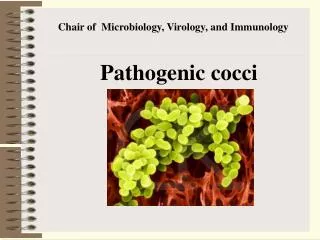
C hair of M icrobiology, V irology, and I mmunology. Pathogenic cocci. Lecturer As. Prof. S.I. Klymnyuk. Staphylococci. Classification. Staphylococci are included in the Firmicutes Bacteria, family Micrococcaceae , genus Staphylococcus .

E N D
Chair of Microbiology, Virology, and Immunology Pathogenic cocci Lecturer As. Prof. S.I. Klymnyuk
Classification. Staphylococci are included in the Firmicutes Bacteria, family Micrococcaceae, genus Staphylococcus. According to the contemporary classification, staphylococci are subdivided into more then 30 species. Among them: S. aureus, S. epidermidis, and S. saprophyticus, S. haemolyticus, S. capitis, S. hominis, S. warneri, S. xylosus etc.
Morphology. Staphylococci are spherical in shape, 0.8-1 mcm in diameter, and form irregular clusters resembling bunches of grapes. In smears from cultures and pus the organisms occur in short chains, in pairs, or as single cocci. Large spherical (L-forms) or very small (G-forms) and even filterable forms may be seen in cultures which have been subjected to various physical, chemical, and biological (antibiotics) factors.
Electron micrograph showing Staphylococcus aureus morphology.
Staphylococci are Gram-positive organisms which possess no flagella and do not form spores.
Cultivation. Staphylococci are facultative-anaerobes. They grow well on ordinary nutrient media with a pH of 7.2-7.4 at a temperature of 37 C but do not grow at temperatures below 10 C and above 45 C. At room temperature with adequate aeration and subdued light – the organisms produce golden, white, lemon-yellow, and other pigments known as lipochromes. These pigments do not dissolve in water but are soluble in ether, benzene, acetone, chloroform, and alcohol.
Cultivation. On meat peptone agar Staphylococci produce well defined colonies with smooth edges, measuring from 1-2 to 2.5 mm in diameter. Growth of Staphylococci in meat-peptone broth produces diffuse opacity throughout the medium and, subsequently, a precipitate. In some cases when there is sufficient aeration, the organisms form a pellicle on the surface of the broth. Staphylococci grow well on potatoes and coagulated serum. After 24-48 hours of incubation there is usually abundant growth along the inoculation stab and liquefaction of gelatin media. On the fourth or fifth day the gelatin medium resembles a funnel filled with fluid. On blood agar pathogenic Staphylococci cause haemolysis of the erythrocytes.
Antigenic structure. Polysaccharide A was extracted from pathogenic strains isolated from patients with septicaemia, furunculosis, osteomyelitis, and acute conjunctivitis, etc. Polysaccharide B is found in avirulent, non-pathogenic strains. Polysaccharides A and B differ not only in their serological reactions but also in their chemical structures. Antigen C, containing a specific polysaccharide, has been recently isolated.
Virulence factors Staphylococci express many cell surface-associated and extracellular proteins that are potential virulence factors. For the majority of diseases caused by this organism, pathogenesis is multifactorial. Thus it is difficult to determine precisely the role of any given factor. This also reflects the inadequacies of many animal models for staphylococcal diseases.
Protein A. Protein A is a surface protein of S aureus which binds immunoglobulin G molecules by the Fc region. In serum, bacteria will bind IgG molecules the wrong way round by this non-immune mechanism. In principle this will disrupt opsonization and phagocytosis. Indeed mutants of S aureus lacking protein A are more efficiently phagocytozed in vitro, and studies with mutants in infection models suggest that protein A enhances virulence.
Leukocidin. S aureus can express a toxin that specifically acts on polymorphonuclear leukocytes. Phagocytosis is an important defense against staphylococcal infection so leukocidin should be a virulence factor.
Membrane Damaging Toxins. a-toxin. The best characterized and most potent membrane-damaging toxin of S aureus is a-toxin. Susceptible cells have a specific receptor for a-toxin which allows low concentrations of toxin to bind, causing small pores through which monovalent cations can pass. At higher concentrations, the toxin reacts non-specifically with membrane lipids, causing larger pores through which divalent cations and small molecules can pass. However, it is doubtful if this is relevant under normal physiological conditions. In humans, platelets and monocytes are particularly sensitive to a-toxin
ß-toxin. ß-toxin is a sphingomyelinase which damages membranes rich in this lipid. The classical test for ß-toxin is lysis of sheep erythrocytes. The majority of human isolates of S aureus do not express ß-toxin. A lysogenic bacteriophage is inserted into the gene that encodes the toxin. This phenomenon is called negative phage conversion. Some of the phages that inactivate the ß-toxin gene carry the determinant for an enterotoxin and staphylokinase.
d-toxin. The d-toxin is a very small peptide toxin produced by most strains of S aureus. It is also produced by S epidermidis and S lugdunensis. The role of d-toxin in disease is unknown. g-toxin and leukocidin. The g-toxin and the leukocidins are two-component protein toxins that damage membranes of susceptible cells. The proteins are expressed separately but act together to damage membranes. There is no evidence that they form multimers prior to insertion into membranes. The g-toxin locus expresses three proteins. The B and C components form a leukotoxin with poor hemolytic activity, whereas the A and B components are hemolytic and weakly leukotoxic.
Superantigens: enterotoxins and toxic shock syndrome toxin. S aureus can express two different types of toxin with superantigen activity, enterotoxins, of which there are six serotypes (A, B, C, D, E and G) and toxic shock syndrome toxin (TSST-1). Enterotoxins cause diarrhea and vomiting when ingested and are responsible for staphylococcal food poisoning. When expressed systemically, enterotoxins can cause toxic shock syndrome (TSS) - indeed enterotoxins B and C cause 50% of non-menstrual TSS.
Epidermolytic (exfoliative) toxin (ET). This toxin causes the scalded skin syndrome in neonates, with widespread blistering and loss of the epidermis and have protease activity. It is not clear how the latter causes epidermal splitting. It is possible that the toxins target a very specific protein which is involved in maintaining the integrity of the epidermis.
Other Extracellular Proteins. Coagulase is an extracellular protein which binds to prothrombin in the host to form a complex called staphylothrombin. The protease activity characteristic of thrombin is activated in the complex, resulting in the conversion of fibrinogen to fibrin. This is the basis of the tube coagulase test, in which a clot is formed in plasma after incubation with the S aureus broth-culture supernatant. Coagulase is a traditional marker for identifying S aureus in the clinical microbiology laboratory.
Staphylokinase.Many strains of S aureus express a plasminogen activator called staphylokinase. The genetic determinant is associated with lysogenic bacteriophages. A complex formed between staphylokinase and plasminogen activates plasmin-like proteolytic activity which causes dissolution of fibrin clots. The mechanism is identical to streptokinase, which is used in medicine to treat patients suffering from coronary thrombosis. As with coagulase there is no evidence that staphylokinase is a virulence factor, although it seems reasonable to imagine that localized fibrinolysis might aid in bacterial spreading.
Enzymes. S aureus can express proteases, a lipase, a deoxyribonuclease (DNase) and a fatty acid modifying enzyme (FAME). The FAME enzyme may be important in abscesses, where it could modify anti-bacterial lipids and prolong bacterial survival. The thermostable DNase is an important diagnostic test for identification of S aureus.
Pathogenicity for animals. Horses, cattle, sheep, goats, pigs, and, among laboratory animals, rabbits, white mice, and kittens are susceptible to pathogenic staphylococci.
Pathogenesis and diseases in man. Staphylococci enter the body through the skin and mucous membranes. When they overcome the lymphatic barrier and penetrate the blood, staphylococcal septicaemia sets in. Both the exotoxins and the bacterial cells play an important role in pathogenesis of diseases caused by these organisms. Consequently, staphylococcal diseases should be regarded as toxinfections. The development of staphylococcal diseases is also influenced by the resulting allergy which in many cases is the cause of severe clinical forms of staphylococcal infections which do not succumb to treatment.
Pathogenesis and diseases May cause infection if the skin or mucous membranes are broken or damaged.Staphylococci are responsible for a number of local lesions in humans: hidradenitis, abscess, paronychia, blepharitis, furuncle, carbuncle, periostitis, osteomyelitis, folliculitis, sycosis,dermatitis, eczema, chronic pyodermia, peritonitis, meningitis, appendicitis,and cholecystitis. Staphylococcus aureus is considered the most pathogenic species, causing abscesses, boils, carbuncles, acne, impetego, and less commonly, pneumonia, osteomyelitis, endocarditis, cystitis, pyelonephritis, and food poisoning. Diabetes mellitus, avitaminosis, alimentary dystrophy, excess perspiration, minor occupational skin abrasions, as well as skinirritation caused by chemical substances, are some examples of theconditions conducive to the formation of pyogenic lesions of the skinand furunculosis.
Pathogenesis and diseases In some cases staphylococci may give rise to secondary infection in individuals suffering from smallpox, influenza, and wounds, as well as postoperative suppurations. Staphylococcal sepsis and staphylococcal pneumonia in children are particularly severe diseases. Ingestion of foodstuffs (cheese, curds, milk, rich cakes and pastry, ice cream, etc.) contaminated with pathogenic staphylococci may result in food poisoning. Staphylococci play an essential part in mixed infections, and are found together with streptococci in cases of wound infections, diphtheria, tuberculosis, actinomycosis, and angina.
Pathogenesis and diseases The wide use of antibacterial agents, antibiotics in particular, led to considerable changes in the severity and degree of the spread of staphylococcal lesions. Growth in the incidence of diseases and intrahospital infections in obstetrical, surgical and children's in-patient institutions, intensive spread of the causative agent, and increase in the number of carriers among the medical staff and population have been noted in all countries of the world. Intrauterine and extrauterine contamination of children with staphylococci has been registered, with the development of vesiculopustular staphyloderma, pemphigus, infiltrates, abscesses, conjunctivitis, nasopharyngitis, otitis, pneumonia, and other diseases.
It has been established that staphylococci become adapted rapidly to chemical agents and antibiotics due to the spread of R-plasmids among these bacteria. The high concentration of drugs in the body of humans and in the biosphere has resulted in essential disturbance in the microflora and the extensive spread of resistant strains possessing more manifest virulence. The L-forms of staphylococci are especially marked by increased degree of resistance to antibiotics.
Immunity. The tendency to run a chronic flaccid course or relapse is regarded as a characteristic symptom of staphylococcal infections. This peculiarity gives a basis for concluding that postinfectional immunity following staphylococcal diseases is of low grade and short duration. Immunity acquired after staphylococcal diseases is due to phagocytosis and the presence of antibodies (antitoxins, precipitins, opsonins, and agglutinins).
Laboratory diagnosis. Test material may be obtained from pus, mucous membrane discharge, sputum, urine, blood, foodstuffs (cheese, curds, milk, pastry, cakes, cream, etc.), vomit, lavage fluids, and faeces. The material is examined for the presence of pathogenic staphylococci. Special rules are observed when collecting the material since non-pathogenic strains are widespread in nature.
Identification of Gram Positive Cocci: Staphylococcus • Contains both pathogenic and non-pathogenic organisms • Do not produce endospores, but are resistant to drying (desiccation) • Found routinely on the surface of the skin • Three major species: • Staphylococcus aureus • Staphylococcus epidermidis • Staphylococcus saprophyticus • The three species can be distinguished from each other by various biochemical tests. • In this lab we will perform some of these tests and observe the results.
Chemical and Biochemical Tests • The identification of organisms is based on cellular, cultural, and biochemical characteristics • All species of Staphylococcus are Gram Positive Cocci (GPC) • On nutrient agar they tend to be white (or cream colored), circular, entire, convex colonies. • On Sheep Blood Agar Staphylococcus aureus may exhibit hemolysis of the agar in the area around the colonies. • Tests to be performed: • Catalase test • Coagulase test • Growth and fermentation on Mannitol Salt Agar • Susceptibility to the antibiotic “Novobiocin”
Catalase Test • The Catalase test determines if the organism produces the enzyme “Catalase”, which breaks down hydrogen peroxide (H2O2) to water and oxygen (O2). Catalase 2 H2O2 2 H2O + O2(g) • Catalase allows organisms to break down harmful metabolites of aerobic respiration and may be seen in aerobic and facultatively anaerobic organisms. There are other enzymes produced by some organisms to handle other toxic end-products of metabolism, such as superoxide dismutase. Not all organisms produce catalase.
Coagulase Test Pathogenic organisms require mechanisms to help them overcome host defense systems. One mechanism involves coating the bacterial cells in a body substance, such as fibrin, to “hide” the bacterial cells from the immune system. This coating will not trigger an immune response by the host cells. The enzyme coagulase causes fibrin to be deposited on bacterial cells helping them to become “invisible” to the host immune system.
High Salt Tolerance • Some organisms cannot tolerate a high salt concentrations. • Media containing higher than normal salt concentrations will inhibit the growth of these non-salt tolerant organisms. • Mannitol salt agar contains a high salt concentrationso only salt tolerant organisms will grow on it. • Also, Mannitol salt agar contains the sugar Mannitol. • Some organisms can utilize this sugar as a food source and will produce acidic by-products from this metabolism. • The addition of acid to the medium by the fermentation of Mannitol changes the pH. • If a pH indicator is present in the medium (such as Phenol red) a color change will occur dependant upon the pH of the medium (agar or broth). • Mannitol Salt Agar contains the pH indicator “Phenol Red” • This pH indicator is red at neutral pH (around 7.0), but turns yellow under acidic conditions.
Antibiotic Susceptibility/Resistance • Antibiotic susceptibility is another test that can be used to identify bacteria. • A paper disc impregnated with the antibiotic, in this case Novobiocin, is placed on a lawn of bacteria following inoculation. • The antibiotic in the disc diffuses into the surrounding agar. • If the bacterial species is susceptible to the antibiotic there is a circle of “no-growth” around the disc where bacterial growth is inhibited by the antibiotic. • If the bacteria is resistant to the antibiotic the cells grow right up the the antibiotic disc. • The bacterial species or strain is reported as being resistant to the antibiotic (R) or susceptible to the antibiotic (S) depending on the observations made. • The diameter of the area of “no-growth” around the disc may determine the susceptibility or resistance of the organism to the antibiotic.
Interpretation of Results Catalase • Bubbling indicates a positive test for the presence of the catalase enzyme. Coagulase • Agglutination of the “Test” latex with no agglutination of the “Control” latex is considered a positive (+) test for the presence of this enzyme. All reactions occurring after 20 seconds should be ignored. • Agglutination of the “Test” latex with no agglutination of the “Control” latex is considered a positive (+) test for the presence of this enzyme.
Mannitol Salt Agar • Two different characteristics of the organism are determined with this agar. The first is the organism’s ability to tolerate a high salt environment. Evidence of growth on the slant indicates the organism can grow in a high salt environment. • Organisms that can ferment the sugar Mannitol produce an acid end-product that changes the red pH indicator (Phenol red) in the media to yellow. • Any yellow in the media is considered a positive test for Mannitol fermentation. • It is possible to have growth, but no Mannitol fermentation.
Novobiocin Susceptibility • A zone of growth inhibition 17 mm or less in diameter indicates resistance (R) to Novobiocin. • If the zone is greater than 11 mm the organism is susceptible (S) to Novobiocin.
Treatment. Staphylococcal diseases are treated with antibiotics (penicillin, phenoxymethyl penicillin, tetracycline, gramicidin, etc.), sulphonamides (norsulphazol, sulphazol, etc.), and antistaphylococcal gamma-globulin.
Prophylaxis. The general precautionary measures include: hygiene in working and everyday-life conditions, treatment of vitamin deficiency, prevention of traumatism and excess perspiration, observance of rules of hygiene in maternity hospitals, surgical departments, children's institutions, industrial plants and enterprises, particularly canneries, observance of personal hygiene and frequent washing of hands in warm water with soap. Routine disinfection of hospital premises (surgical departments, maternity wards) and bacteriological examination of the personnel for carriers of pathogenic staphylococci resistant to antibiotics are also necessary. To prevent pyoderma protective ointments and pastes are used at industrial enterprises. In some cases specific prophylaxis by means of immunization with the staphylococcal anatoxin may be recommended for individuals subject to injury or infection with antibiotic-resistant staphylococci.