Download
1 / 65
650 likes | 794 Views
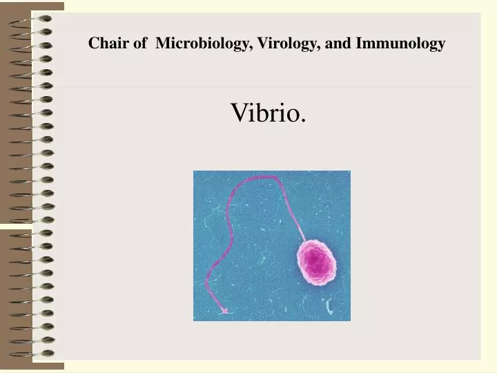
C hair of M icrobiology, V irology, and I mmunology. Vibrio. Causative agents of Shigellosis.

E N D
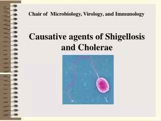
Causative agents of Shigellosis The causative agent of dysentery was described in 1888 by A. Chantemesse and in 1891 by A. Grigoryev and F. Widal. In 1898 this organism was studied in detail by K. Shiga in Japan and in 1900-1901 by V. Kruse in Germany (Shiga bacillus).
Morphology. Morphologically dysentery bacilli correspond to the organisms of the family Enterobacteriaceae. Dysentery bacilli have no flagella and this is one of the differential characters between these organisms and bacteria of the coli-typhoid-paratyphoid group. Some strains of Flexner bacilli are found to possess cilia.
Toxin production. S. dysenteriae produce thermolabile exotoxin which displays marked tropism to the nervous system and intestinal mucous membrane. This toxin may be found in old meat broth cultures, lysates of a 24-hour-old agar culture, and in desiccated bacterial cells.
The dysentery exotoxin causes the production of a corresponding antitoxin. The remaining types of dysentery bacilli produce no soluble toxins. They contain endotoxins, which are of a gluco-lipo-protein nature, and occur in the smooth but not in the rough variants. Thermolabile substances exerting a neurotropic effect were revealed in some S. sonnei strains. They were extracted from old cultures by treating the latter with trichloracetic acid.
Epidemiology and Pathogenesis of Shigellosis. Humans seem to be the only natural hosts for the shigellae, becoming infected after the ingestion of contaminated food or water. Unlike Salmonella, the shigellae remain localized in the intestinal epithelial cells, and the debilitating effects of shigellosis are mostly attributed to the loss of fluids, electrolytes, and nutrients and to the ulceration that occurs in the colon wall. It has been known for many years that Shigella dysenteriae type 1 secreted one or more exotoxins (called Shiga toxins), which would cause death when injected into experimental animals and fluid accumulation when placed in ligated segments of rabbit ileum.
Laboratory diagnosis. Reliable results of laboratory examination depend, to a large extent, on correct sampling of stool specimens and its immediate inoculation onto a selective differential medium. The procedure should be carried out at the patient's bedside, and the plate sent to the laboratory.
For the serological diagnosis of dysentery the indirect haemagglutination (IHA) test with erythrocyte diagnosticums with the titre of 1:160 and higher is performed. The test. is repeated after at least seven days. Diagnostically important is a four-fold rise in the antibody litre, which can be elicited from the 10th-12th day of the disease. To distinguish between patients with subclinical forms of the disease and Shigella carriers, identify immunoglobulins of the G class.
Treatment and Control of Shigellosis Intravenous replacement of fluids and electrolytes plus antibiotic therapy are used for severe cases of shigellosis. Ampicillin frequently is not effective, and alternative therapies include sulfamethoxazole/trimethoprim and, with increasing sulfamethoxazole/trimethoprim resistance, the quinolone antibiotics such as nalidixic acid and ciprofloxacin. In the Far East, India, and Brazil where shigellosis is more common than in the United States, multiple antibiotic resistance because of the acquisition of plasmids has become common. Shigellosis also is common in Latin America.
The injection of killed vaccines is worthless, because humoral IgG does not seem to be involved in immunity to the localized intestinal infection. Live vaccines that cannot grow in the absence of streptomycin (ie, streptomycin-dependent vaccines) have been developed and used in clinical trials, but success has been equivocal.
Summary SHIGELLA 1. Genus characteristics a. Slender, gram-negative rod; non lactose-fermenting (except for S. sonnei) b. In contrast to E. coli: no H2S production, no lysine decarboxylation, no acetate utilization c. Invasive (key to pathogenesis) d. In contrast to Salmonella: non-motile; no gas from glucose fermentation; no H2S production e
e. Toxin production limited to a few strains f. All have O antigens-four groups (A-D) g. Differentiating species i. S. dysenteriae - no mannitol fermentation ii. S. boydii - C antigen group iii. S. flexneri - B antigen group iv. S. sonnei-orniltine decarbexylase production
h. Specimens i. Rectal swab from colonic ulcer is best for culture. ii. Fecal specimen - must be immediately innoculated onto transport media or culture media.Sensitive to acids present in feces.
2. Epidemiologya. Species i. S. sonnei most frequent isolate in U.S.ii. S. dysenteriae and S. boydii most frequently isolated in developing countries - cause moresevere disease course. iii. S. flexneri more commonly isolated from homosexual menb. Humans and higher primates are only natural reservoir.c. High communicability (< 200 bacilli needed to produce disease)d. May be spread by fomites;”food, fingers, feces, and files”.
3. Clinical manifestations a. Desease - bacillary dysentery (shigellosis), characterized by painful, frequent, low-volume stools; feces contain blood and mucous. Bacteremia is rare since organisms usually do not enter bloodstream. Polymorphonuclear leukocyles present in stool.b. Carriers i. One to four weeks after the disease; carrier state may be set up if organism is not cleared.ii. Long-term; recurrent bouts of disease
4. Therapy and prevention a. Hydration and electrolyte replacementb. Ampicillin - decreases duration of symptoms and carrier state; tetracycline; trimethoprim-sulfamelhoxazolec. Prevention through personal hygiene, proper garbage disposal and water purification.
The causative agents of cholera are the classical Vibrio cholera biovars discovered by R. Koch in 1883 and the El Tor vibrio biovar isolated from the cadaver of a pilgrim on the Sinai peninsula by Gotschlich in 1906. Vibrio cholerae biovar Proteus (N. Gamaleya, 1888) and Vibrio cholerae biovar albensis were discovered" later. V. cholerae was described by F. Pacini in 1854.
Classification. Vibrio cholerae belongs to family Vibrionaceae, genus Vibrio consisting of 5 species. The species Vibrio cholerae is subdivided into four biological variants: biovar cholerae, biovar El Tor, biovar Proteus, and biovar albensis. Biovar cholerae and biovar El Tor of Vibrio cholerae are the causative agents of human cholera. Biovar Proteus of Vibrio cholerae causes diarrhoea in birds and gastroenteritis in humans; biovar albensis of Vibrio cholerae was revealed in fresh water and in human faeces and bile.
Morphology. Cholera vibrios are shaped like a comma or a curved rod measuring 1-5 mcm in length and 0.3 mcm in breadth They are very actively motile, monotrichous, nonsporeforming, noncapsulated, and Gram-negative.
Cultivation. Cholera vibrios are facultative (anaerobes). The optimum growth temperature is 37° C, and growth is arrested below 14 °C and above 42° C. The organisms grow readily on alkaline media at pH 6.0-9.0, and on solid media the colonies are transparent with a light-blue hue, forming domes with smooth edges.
Toxin production. The cholera vibrio produces an exotoxin (cholerogen) which is marked by an enterotoxic effect and plays an important role in the pathogenesis of cholera; the endotoxin also exerts a powerful toxic effect. The cholera vibrios produce fibrinolysin, hyaluronidase, collagenase, mucinase, lecithinase, neuraminidase, and proteinases.
Remember that non-O1 and non-O139 strains of V.cholerae also cause a wide spectrum of infections, ranging from mild diarrhea to one indistinguishable from classic cholera. Some of these serotypes are known to produce a choleratoxin that is identical to that of the classic biotypes, whereas other products a heat-stable enterotoxin analogous to the ST of E. coli.
Antigenic structure. The cholera vibrios have thermostable O-antigens (somatic) and thermolabile H-antigens (flagellar). The O-antigen possesses species and type specificity, the H-antigen is common for the genus Vibrio. The cholerae vibrios, El Tor biovars and biovars cholera belong to the O-1 subgroup.
Pathogenesis and diseases in man. Cholera is undoubtedly the most dramatic of the water-borne diseases. As far as is known, cholera was confined to India for the almost 2000 years between its first description by Hindu physicians in 400 b c and its spread to Arabia, Persia, Turkey, and Southern Russia in the early1800s. There were six major pandemics of cholera during the 1800s covering the entire world, killing millions wherever it struck.
Three phases can be distinguished in the development of the disease. 1. Cholera enteritis (choleric diarrhoea) which lasts 1 or 2 days. 2.Cholera gastroenteritis. Profuse diarrhoea and continuous vomiting lead to dehydration of the patient's body and this results in lowering of body temperature, decrease in the amount of urine excreted, drastic decrease in the number of mineral and protein substance, and the appearance of convulsions. The presence of cholera vibrios is revealed guite frequently in 3.Cholera algid which is characterized by severe symptoms. The skin becomes wrinkled due to the loss of water, cyanosis appears, and the voice becomes husky and is sometimes lost completely. The body temperature falls to 35.5-34 °C. As a result of blood concentration cardiac activity is drastically weakened and urination is suppressed.
Laboratory diagnosis. A strict regimen is established in the laboratory. Examinations are carried out in accordance with the general rules observed for particularly hazardous diseases. Test specimens are collected from stools, vomit, organs obtained at autopsy, water, objects contaminated by patient's stools, and, in some cases, from foodstuffs. Certain rules are observed when the material is collected and transported to the laboratory, and examination is made in the following stages.
1. Stool smears stained by a water solution of fuchsin are examined microscopically. In the smears, the cholera vibrios occur in groups similar to shoals of fish (Fig. 3). 2. A stool sample is inoculated into 1 per cent peptone water and alkaline agar. After 6 hours incubation at 37°C the cholera vibrios form a thin pellicle in the peptone water, which adheres to the glass. The pellicle smears are Gram stained, and the culture is examined for motility. A slide agglutination reaction is performed with specific agglutinating O-serum diluted in a ratio of 1 in 100.
The organisms are then transferred from the peptone water onto alkaline agar for isolation of the pure culture. If the first generation of the vibrios in peptone water is not visible, a drop taken from the surface layer is re-inoculated into another tube of peptone water. In some cases with such re-inoculations, an increase in the number of vibrios is achieved. The vibrio culture grown on solid media is examined for motility and agglutinable properties. Then it is subcultured on an agar slant to obtain the pure culture.
3. The organism is identified finally by its agglutination reaction with specific O-serum, determination of its fermentative properties (fermentation of mannose, saccharose, and arabinose), and its susceptibility to phagolysis
Treatment. The mortality rate of cholera can be reduced to less than1% by the adequate replacement of fluids and electrolytes. Antibiotics of the tetracycline group (tetracycline, sigmamycin), amphenicol, and streptomycin are prescribed at first intravenously and then by mouth. Pathogenetic therapy is very important: control of dehydration, hypoproteinaemia, metabolic disorders, and the consequences of toxicosis, acidosis in particular, by infusion of saline (sodium and potassium) solutions, infusion of plasma or dry serum, glucose, the use of warm bath, administration of drugs which improve the tone of the heart and vessels.
Prophylaxis. The following measures are applied in a cholera focus: (1) detection of the first cases with cholera, careful registration of all sick individuals, immediate information of health protection organs; 2) isolation and hospitalization, according to special rules, of all sick individuals and carriers, observation and laboratory testing of all contacts; (3) concurrent and final disinfection in departments for cholera patients and in the focus;
(4) protection of sources of water supply, stricter sanitary control over catering establishments, control of flies; in view of the possibility of El Tor vibrio reproducing in water reservoirs under favourable conditions (temperature, the presence of nutrient substrates) systematic bacteriological control over water reservoirs has become necessary, especially in places of mass rest and recreation of the population in the summer;
(5) strict observance of individual hygiene; boiling or proper chlorination of water, decontamination of dishes, hand washing; (6) specific prophylaxis: immunization with the cholera monovaccine containing 8 thousand million microbial bodies per 1 ml or with the cholera anatoxin. Chemoprophylaxis with oral tetracycline is conducted for persons who were in contact with the sick individual or for patients with suspected cholera.
SUMMURY I. GENUS VIBRIO A. GENERAL CHARACTERISTICS 1. From family Vibnonaceae2 Naturally occur in water-marine and fresh waters; some occur in cold-blooded animals. 3. Oxidase-positive-differentiates Vibrios from Enterobacteriaceae. 4. Characteristic comma shape
B VIBRIO CHOLERAE 1 Species characteristics a. Comma-shaped when first isolated b. Aerobic c. Unipolar flagellum-motile d. Primary isolation-simple media, MacConke/s agar, tellurite taurocholate gelatin agar (TTGA), thiosulfate citrate bile salts agar (TCBS)e. Sensitive to 2,4-diamme-6,7 d isopropyi pteridine (0/129), useful in distinguishing from other gram-negative, oxidase-posilive bacilli (i.e., Aeromonas)f. Prefer alkaline environment.