Download
1 / 35
440 likes | 1.17k Views
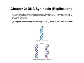
Generalized model of semiconservative replication of DNA. New synthesis is shown in teal. Results of one round of replication of DNA for each of the three possible modes by which replication could be accomplished. The Meselson-Stahl experiment.

E N D
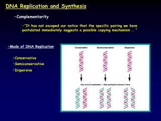
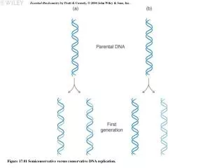
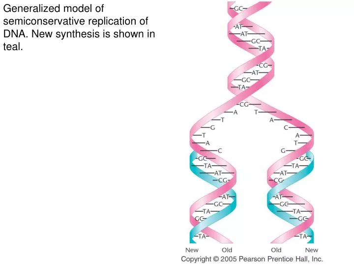
Generalized model of semiconservative replication of DNA. New synthesis is shown in teal.
Results of one round of replication of DNA for each of the three possible modes by which replication could be accomplished.
The expected results of two generations of semiconservative replication in the Meselson-Stahl experiment.
The Taylor-Woods-Hughes experiment, demonstrating the semiconservative mode of replication of DNA in root tips of Vicia faba. (a) An unlabeled chromosome proceeds through the cell cycle in the presence of 3H-thymidine. As it enters mitosis, both sister chromatids of the chromosome are labeled, as shown by autoradiography. After a second round of replication (b), this time in the absence of 3H-thymidine only one chromatid of each chromosome is expected to be surrounded by grains. Except where a reciprocal exchange has occurred between sister chromatids (c), the expectation was upheld.
Bidirectional replication of the E. coli chromosome. The thin black arrows identify the advancing replication forks.
The chemical reaction catalyzed by DNA polymerase I. During each step, a single nucleotide is added to the growing complement of the DNA template using a nucleoside triphosphate as the substrate. The release of inorganic pyrophosphate drives the reaction energetically.
Helical unwinding of DNA during replication as accomplished by DnaA, DnaB, and DnaC proteins. Initial binding of many monomers of DnaA occurs at DNA sites that contain repeating sequences of nine nucleotides, called 9mers. Not illustrated are 13mers, which are also involved.
The initiation of DNA synthesis. A complementary RNA primer is first synthesized, to which DNA is added. All synthesis is in the 5’-to-3’ direction. Eventually, the RNA primer is replaced with DNA under the direction of DNA polymerase I.
Opposite polarity of DNA synthesis along the two strands, necessary because the two strands of DNA run antiparallel to one another and DNA polymerase III synthesizes only in one direction (5’-to-3’). On the lagging strand, synthesis must be discontinuous, resulting in the production of Okazaki fragments. On the leading strand, synthesis is continuous. RNA primers are used to initiate synthesis on both strands.
Illustration of how concurrent DNA synthesis may be achieved on both the leading and lagging strands at a single replication fork. The lagging template strand is “looped” in order to invert the physical direction of synthesis, but not the biochemical direction. The enzyme functions as a dimer, with each core enzyme achieving synthesis on one or the other strand.
A demonstration of the multiple origins of replication along a eukaryotic chromosome. Each origin is apparent as a replication bubble along the axis of the chromosome. Arrows identify some of these.
An electron micrograph of a eukaryotic-replicating fork demonstrating the presence of histone protein-containing nucleosomes on both branches.
The difficulty encountered during the replication of the ends of linear chromosomes: A gap (marked --b--) cannot be filled following synthesis on the lagging strand.
The predicted solution to the difficulty posed in Figure 11-16. The enzyme telomerase directs synthesis of the TTGGGG sequences, which results in the formation of a hairpin structure. The gap can now be filled, and the hairpin structure is cleaved.
Model depicting how genetic recombination can occur as a result of the breakage and rejoining of heterologous DNA strands. Each stage is described in the text.
The electron micrograph shows DNA in a chi-form structure similar to the diagram in (g); the DNA is an extended Holliday structure, derived from the ColE1 plasmid of E. coli