Download
1 / 25
261 likes | 494 Views
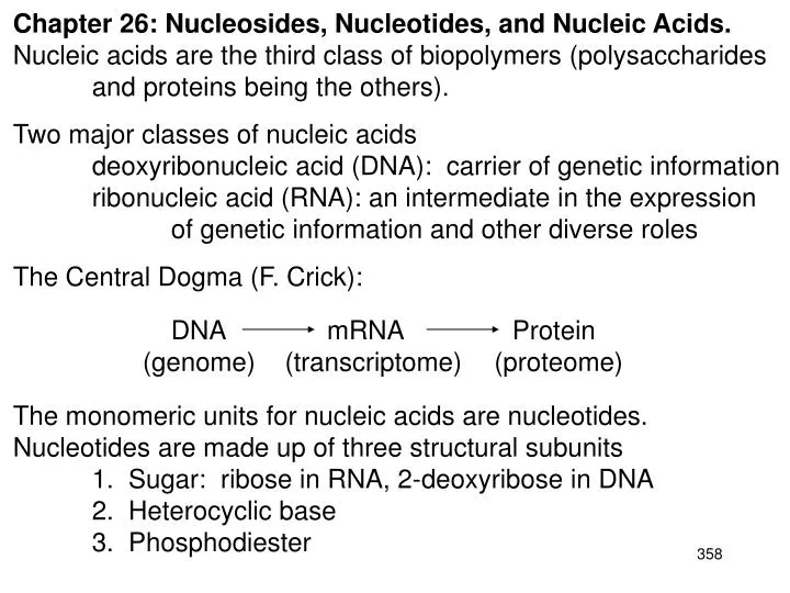
Chapter 26: Nucleosides, Nucleotides, and Nucleic Acids. Nucleic acids are the third class of biopolymers (polysaccharides and proteins being the others). Two major classes of nucleic acids deoxyribonucleic acid (DNA): carrier of genetic information

E N D
Chapter 26: Nucleosides, Nucleotides, and Nucleic Acids. Nucleic acids are the third class of biopolymers (polysaccharides and proteins being the others). Two major classes of nucleic acids deoxyribonucleic acid (DNA): carrier of genetic information ribonucleic acid (RNA): an intermediate in the expression of genetic information and other diverse roles The Central Dogma (F. Crick): DNA mRNA Protein (genome) (transcriptome) (proteome) The monomeric units for nucleic acids are nucleotides. Nucleotides are made up of three structural subunits 1. Sugar: ribose in RNA, 2-deoxyribose in DNA 2. Heterocyclic base 3. Phosphodiester
26.1: Pyrimidines and Purines. The heterocyclic bases; there are six common bases for nucleic acids (Table 26.1, p. 1177). Note that G, T and U exist in the keto form (and not the enol form found in phenols) 26.2: Nucleosides. N-Glycosides of a purine or pyrimidine heterocyclic base and a carbohydrate. The C-N bond involves the anomeric carbon of the carbohydrate. The carbohydrates for nucleic acids are D-ribose and 2-deoxy-D-ribose
Nucleosides = carbohydrate + base (Table 28.2, p. 1168) ribonucleosides or 2’-deoxyribonucleosides To differentiate the atoms of the carbohydrate from the base, the position number of the carbohydrate is followed by a ´ (prime). The stereochemistry of the glycosidic bond found in nucleic acids is .
26.3: Nucleotides. Phosphoric acid esters of nucleosides. Nucleotides = nucleoside + phosphate Kinase: enzymes that catalyze the phosphoryl transfer reaction from ATP to an acceptor substrate. M2+ dependent
1971 Nobel Prize in Medicine or Physiology: Earl Sutherland 26.4: Bioenergetics. (Please read) 26.5: ATP and Bioenergetics. (Please read)
26.6: Phosphodiesters, Oligonucleotides, and Polynucleotides. The chemical linkage between nucleotide units of nucleic acids is a phosphodiester, which connects the 5’-hydroxyl group of one nucleotide to the 3’-hydroxyl group of the next nucleotide. By convention, nucleic acid sequences are written from left to right, from the 5-end to the 3’-end. Nucleic acids are negatively charged 5’-direction RNA, X= OH DNA, X=H 3’-direction
26.7: Nucleic Acids (please read). Chargaff’s Rule: A=T and C=G 26.8: Secondary Structure of DNA: The Double Helix. Two polynucleotide strands, running in opposite directions (anti-parallel) and coiled around each other in a double helix. The strands are held together by complementary hydrogen- bonding between specific pairs of bases. (Fig. 26.2, p. 1187)
DNA double helix major groove 12 Å one helical turn 34 Å minor groove 6 Å backbone: deoxyribose and phosphodiester linkage bases (Figs. 26.4 & 26.5, p. 1189)
26.9: Tertiary Structure of DNA: Supercoils. Each cell contains about two meters of DNA. DNA is “packaged” by coiling around a core of proteins known as histones. The DNA-histone assembly is called a nucleosome. Histones are rich is lysine and arginine residues. Pdb code 1kx5
“It has not escaped our attention that the specific pairing we have postulated immediately suggests a possible copying mechanism for the genetic material.” Watson & Crick 26.10: Replication of DNA. The Central Dogma (F. Crick): DNA replication DNA transcription mRNA translation Protein (genome) (transcriptome) (proteome) Expression and transfer of genetic information: Replication: process by which DNA is copied with very high fidelity. Transcription: process by which the DNA genetic code is read and transferred to messenger RNA (mRNA). This is an intermediate step in protein expression Translation: The process by which the genetic code is converted to a protein, the end product of gene expression. The DNA sequence codes for the mRNA sequence, which codes for the protein sequence
DNA is replicated by the coordinated efforts of a number of proteins and enzymes. For replication, DNA must be unknotted, uncoiled and the double helix unwound. Topoisomerase: Enzyme that unknots and uncoils DNA Helicase: Protein that unwinds the DNA double helix. DNA polymerase: Enzyme that replicates DNA using each strand as a template for the newly synthesized strand. DNA ligase: enzyme thatcatalyzes the formation of the phosphodiester bond between pieces of DNA. DNA replication is semi-conservative: Each new strand of DNA contains one parental (old, template) strand and one daughter (newly synthesized) strand
Unwinding of DNA by helicases expose the DNA bases (replication fork) so that replication can take place. Helicase hydrolyzes ATP in order to break the hydrogen bonds between DNA strands DNA replication (Fig. 26.8, p. 1192) animation: http://www.hhmi.org/biointeractive/dna/DNAi_replication_vo2.html
DNA Polymerase: the new strand is replicated from the 5’ 3’ (start from the 3’-end of the template) DNA polymerases are Mg2+ ion dependent The deoxynucleotide 5’-triphosphate (dNTP) is the reagent for nucleotide incorporation dNTP (Fig 26.9, p. 1193) 3’-hydroxyl group of the growing DNA strand acts as a nucleophile and attacks the -phosphorus atom of the dNTP.
Replication of the leading strand occurs continuously in the 5’ 3’ direction of the new strand. Replication of the lagging strand occurs discontinuously. Short DNA fragments are initially synthesized and then ligated together. DNA ligase catalyzes the formation of the phosphodiester bond between pieces of DNA. (Fig. 26.8, p. 1192)
26.11 Ribonucleic Acid RNA contains ribose rather than 2-deoxyribose and uracil rather than thymine. RNA usually exist as a single strand. There are three major kinds of RNA messenger RNA (mRNA): ribosomal RNA (rRNA) transfer RNA (tRNA) DNA is found in the cell nucleus and mitochondria; RNA is more disperse in the cell.
Transcription: only one of the DNA strands is copied (coding or antisense strand). An RNA polymerase replicates the DNA sequence into a complementary sequence of mRNA (template or sense strand). mRNAs are transported from the nucleus to the cytoplasm, where they acts as the template for protein biosynthesis (translation). A three base segment of mRNA (codon) codes for an amino acid.The reading frame of the codons is defined by the start and stop codons. (Table 26.4, p. 1194)
The mRNA is positioned in the ribosome through complementary pairing of the 5’-untranslated region of mRNA with a rRNA. Transfer RNA (tRNA): t-RNAs carries an amino acid on the 3’-terminal hydroxyl (A) (aminoacyl t-RNA) and the ribosome catalyzes amide bond formation. Ribosome: large assembly of proteins and rRNAs that catalyzes protein and peptide biosynthesis using specific, complementary, anti-parallel pairing interactions between mRNA and the anti-codon loop of specific tRNA’s.
Although single-stranded, there are complementary sequences within tRNA that give it a defined conformation. The three base codon sequence of mRNA are complementary to the “anti-codon” loops of the appropriate tRNA. The base- pairing between the mRNA and the tRNA positions the tRNAs for amino acid transfer to the growing peptide chain. variable loop TC loop D loop aminoacyl t-RNA (Fig. 26.11, p. 1195)
E site 26.12: Protein Biosynthesis. Ribosomal protein synthesis A site P site (Fig. 26.12, p. 1196) animation: http://www.hhmi.org/biointeractive/dna/DNAi_translation_vo2.html
26.13: AIDS. (please read) 26.14: DNA Sequencing. Maxam-Gilbert: relies on reagents that react with a specific DNA base that can subsequent give rise to a sequence specific cleavage of DNA Sanger: Enzymatic replication of the DNA fragment to be sequenced with a DNA polymerase, Mg+2, and dideoxynucleotides triphosphate (ddNTP) that truncates DNA replication Restriction endonucleases: Bacterial enzymes that cleave DNA at specific sequences 5’-d(G-A-A-T-T-C)-3’ 3’-d(C-T-T-A-A-G)-5’ 5’-d(G-G-A-T-C-C)-3’ 3’-d(C-C-T-A-G-G)-5’ EcoR I BAM HI
Sanger Sequencing: key reagent:dideoxynucleotides triphosphates (ddNTP) When a ddNTP is incorporated elongation of the primer is terminated The ddNTP is specifically incorporated opposite its complementary nucleotide base
Sanger Sequencing Larger fragments Smaller fragments http://www.hhmi.org/biointeractive/dna/DNAi_sanger_sequencing.html
26.15: The Human Genome Project. (please read) 26.16: DNA Profiling and Polymerase Chain Reaction (PCR). method for amplifying DNA using a DNA polymerase, dNTPs and cycling the temperature. Heat stable DNA Polymerases (from archaea): Taq: thermophilic bacteria (hot springs)- no proof reading Pfu: geothermic vent bacteria- proof reading Mg 2+ two Primer DNA strands (synthetic, large excess) one sense primer and one antisense primer one Template DNA strand (double strand) dNTP’s 1 x 2 = 2 x 2 = 4 x 2 = 8 x 2 = 16 x 2 = 32 x 2 = 64 x 2 = 128 x 2 = 256 x 2 = 512 x 2 = 1,024 x 2 = 2,048 x 2 = 4,096 x 2 = 8,192 x 2 = 16,384 x 2 = 32,768 x 2 = 65,536 x 2 = 131,072 x 2 = 262,144 x 2 = 524,288 x 2 = 1,048,576 In principle, over one million copies per original, can be obtained after just twenty cycles KARY B. MULLIS, 1993 Nobel Prize in Chemistry for his invention of the polymerase chain reaction (PCR) method.
Polymerase Chain Reaction (Fig. 26.14, p. 1202-3) For a PCR animation go to: http://www.youtube.com/watch?v=ZmqqRPISg0g