Download
1 / 50
500 likes | 661 Views
A Case Study of a Patient with Brain Tumor Presented by: Beena Babu. I. DEMOGRAPHIC DATA. Name: Patient X Age: 76years old Sex: Male Nationality: Saudi Date of Admission: 27/09/2012 Diagnosis : Brain Tumour. II. PHYSICAL ASSESSMENT.

E N D
A Case Study of a Patient with Brain TumorPresented by: BeenaBabu
I. DEMOGRAPHIC DATA • Name: Patient X • Age: 76years old • Sex: Male • Nationality: Saudi • Date of Admission: 27/09/2012 • Diagnosis : Brain Tumour
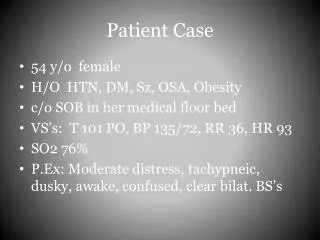
II. PHYSICAL ASSESSMENT Central Nervous System • Upon admission LOC: Vegetative state, GCS: Eye-4 Verbal-1 Motor-1 = 6. Pupil Size of 2-3mm (both pupils), Language and communication: unable to talk , not able to follow simple commands. Not responsive to both verbal and physical stimuli, positive seizure episode.
Cardiovascular System • Vital signs: BP: 101/68mmHg, HR: 72bpm, T: 36.4 degree Celsius, O2 sat 96-98 %. With no neck vein enlargement, Pink mucus membrane noted, (-) heart murmurs, Capillary Refill: 2-3 secs. Full and equal peripheral pulses, No edema noted, No cyanosis.
Respiratory System • Symmetric chest expansion, no retractions, clear breath sounds, No nasal flaring • . . Breathing pattern is regular and not in respiratory distress. With mild white Secretions noted upon suctioning.
Gastrointestinal System • With nasogastric tube inserted on right nostril intact and patent, Flabby, soft, non-tender abdomen, With normoactive abdominal bowel sounds (15 per minute). On Ensure plus 200ml plus 100ml of water per flushing.. Bowel pattern of every other day or every after 3 days (fleet enema). Stool is semi solid in moderate amount. No abdominal pain
Urinary System • He was on foleys catheter before and currently on condom catheter via urine bag draining well with straw colour urine output. No bladder distension with Urine output of 1,400 – 1,900ml in 24 hours. No urgency nor dysuria noted.
Integumentary System • Fair in complexion with good skin turgor, with a wrinkled skin due to loss of elastic fiber and decreased subcutaneous fat from hypodermis secondary to aging. skin integrity is intact, dry and warm skin. With no active dermatosis, no evidence of impending decubitus formation noted. No edema and No IV access. With bedsores on both buttocks stage 3 which means full thickness skin loss involving damage or necrosis of subcutaneous tissue, which may extend down to but not through underlying fascia. The ulcer presents clinically as a deep crater with or without undermining of adjacent tissue. Tunneling, granulating, epithelization and exudates purulent discharge. Length of 3.1cm, width of 3cm and depth of 0.5cm
Musculoskeletal System • Patient is on complete bed rest, Turning is done every 2 hours withHours of sleep: 2-3 hours, interrupted during feeding.. unable to move with Muscle Strength of 1/5( both upper extremities and both lower extremities muscle contraction is noted but no movement occurs. The muscle is not strong enough to lift the particular body part against gravity or move it when in a gravity reduced position). unable to turn side-to-side
III. PATIENT HISTORY Past Medical History • Patient X who have been known to have DM,HTN,braintumor in right temporal area with craniotomy on chemotherapy treatment was shifted from RMH to AAHon 28/06/2012 for long term treatment.On admission patient is receving food and medicine orally.
Present Medical History Patient X is now admitted here in Dr.Abanamy Hospital for long term nursing care and management. Currently, he is on NGT (naso gastric tube).On admission patient was conscious,GSC 13/15 ,disoriented.His vital signs are stable.He is on cap.phenytion 200mg bid,clonazepam,atenolol,NPH insulin. Radiation completed.Due to general weakness and detoriation chemotherapy was discontinued.
Past surgical history: • Patient undergone craniotomy on16/09/2012, due to epidural hematoma.
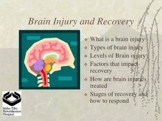
IV. TOPIC PRESENTATION • BRAIN TUMOR • It is a abnormal proliferation of cells in the central nervous system.A tumour is a mass of cancerous cells with in the brain. • Brain tumors include all tumors inside the cranium or in the central spinal canal. They are created by an abnormal and uncontrolled cell division, usually in the brain itself, but also in lymphatic tissue, in blood vessels, in the cranial nerves, in the brain envelopes (meninges), skull, pituitary gland, or pineal gland. Within the brain itself, the involved cells may be neurons or glial cells (which include astrocytes, oligodendrocytes, and ependymal cells). Brain tumors may also spread from cancers primarily located in other organs (metastatic tumor.
The cranium • The brain is protected by a bony covering called the cranium (which, along with the bones of the face, make up the skull). Inside the cranium, the brain is surrounded by the meninges. The meninges is made up of 3 layers of tissue
The cerebrum The largest part of the brain located in the front. It is responsible for movement, body temperature, touch, vision, hearing, judgment, reasoning, problem solving, emotions and learning.
The brainstem • The brainstem is located in front of the cerebellum in he middle of the brain. It is the main control panel for the body that passes messages back and forth between the brain and other parts of the body. The cerebrum, the cerebellum, and the spinal cord are all connected to the brainstem.
The cerebellum • Behind the cerebrum at the back of the head is the cerebellum. The cerebellum is primarily a movement control center, responsible for voluntary muscle movements, fine motor skills, maintaining balance, posture, and equilibrium. Unlike the cerebrum, the
Cranial nerves • The brain also contains 12 pairs of cranial nerves each responsible for specific functions in the body: • Olfactory nerve – smell • Optic nerve – vision • Oculomotor – eye movements, eyelid opening • Trochlear – eye movements • Trigeminal – facial sensations, chewing • Abducens – eye movements • Facial – taste, facial expressions • Vestibulocochlear – hearing, balance • Glossopharyngeal – taste, swallowing • Vagus – swallowing, taste • Accessory – neck and shoulder muscles • Hypoglossal – tongue movement
Medulla Oblongata • Located above spinal cord. It regulates vital functions, such as heartbeat and breathing. The Medulla Oblongata is responsible for coughing, sneezing, vomiting, salivating, swallowing, gaging
Spinal Cord This structure is responsible for basic vital life functions such as breathing, heartbeat, and blood pressure.
Focal signs and symptoms Anterior frontal lobe • Long/short term memory loss • Difficulty in problem solving/concentrating and calculating • Slowness of reaction • Behavioural changes • Emotional liability • Loss of social behaviour
Posterior frontal lobe • Fluent speech deficit • Difficulty with word findings • Motor weakness • Focal seizure activity
Parietal lobe • Deficit in sensation • Inability to recognise common object/numbers/letters Temporal lobe • Psychomotor deficits • Weakness • Seizures • Visual field deficit • Memory deficit • Hallucination • Amnesia
Occipital lobe • Visual deficit • Visual hallucination • Unable to follow commands Hypothalamus region • Visual deficit • Head ache • Acromegaly • hypopituitarism
lateral and third ventricle • head ache • nausea • vomiting • increase ICP Fourth ventricle • head ache • nausea • vomiting • increase ICP
Risk factors for brain tumour • ionizing radiation: from high dose x-ray(radiation therapy)cause cell damage. • Family history: people with various inherited diseases such as multiple endocrine neoplasia,neurofibromatosis are high risk of developing brain tumour.
Aside from exposure to vinyl chloride or ionizing radiation, there are no known environmental factors associated with brain tumors. Mutations and deletions of so-called tumor suppressor genes are thought to be the cause of some forms of brain tumors. People with various inherited diseases, such as Von Hippel-Lindau syndrome, multiple endocrine neoplasia, neurofibromatosis type 2 are at high risk of developing brain tumors. • Although studies have not shown any link between cell phone radiation and brain tumors,[4] the World Health Organization has classified mobile phone radiation on the IARC scale into Group 2B – possibly carcinogenic. That means that there "could be some risk" of carcinogenicity.
PATHOPHYSIOLOGY • Due to etiological factor • Like chemical and radiation Exposure. • Irritation and damage to brain cells • Changes in cell morphology • DNA gene mutation • Inactivation of tumor suppressor and activation of oncogenes • Un controlled cell division and decreased apoptosis. • Hyperplasia of the brain cells • Brain tumour.
TYPES • Tumors can be benign or malignant, can occur in different parts of the brain, and may or may not be primary tumors. A primary tumor is one that has started in the brain, as opposed to a metastatictumor, which is something that has spread to the brain from another part of the body.[6] The incidence of metastatic tumors are more prevalent than primary tumors by 4:1. • [7]Tumors patient has symptoms, others show up incidentally on an imaging scan, or at an autopsy.
The most common primary brain tumors are: • Gliomas(50.4%) • Meningiomas(20.8%) • Pituitary adenomas(15%) • Nerve sheath tumors(8%) • BrainTumor are classified as grade 1,2,3and4. Most common type of primary brain tumours among adults are menigioma and astrocytoma. • For children Meduloblastoma grade 1 or 2 ,astrocytoma,ependymoma.
Signs and Symptoms The symptoms of brain tumors depend on their size and location in the brain. Symptoms often are caused by damage to vital tissue and pressure on the brain as the tumor grows within the limited space in the skull. They may be caused by swelling and a buildup of fluid around the tumor, a condition called edema. Symptoms also may be due to hydrocephalus, which occurs when the tumor blocks the flow of cerebrospinal fluid and causes a build-up in the ventricles. • If a brain tumor grows very slowly, its symptoms may not appear for some time. The most frequent symptoms of brain tumors include: • Headaches that tend to be worse in the morning and ease during the day • Seizures or convulsions • Nausea or vomiting • Weakness or loss of feeling in the arms or legs • Stumbling or lack of coordination in walking • Abnormal eye movements or changes in vision • Drowsiness • Changes in personality or memory
Changes in speech • Double vision • Hearing loss • These symptoms may be caused by brain tumors or by other problems. Diagnostic tests can be performed to determine if the cause of your symptoms is a brain tumor and if it is a primary or secondary one .
Treatment • When a brain tumor is diagnosed, a medical term will be formed to assess the treatment options presented by the leading surgeon to the patient and his/her family. These various types of treatment are available depending on neoplasm type and location and may be combined to give the best chances of survival: • Surgery:complete or partial resection of the tumor with the objective of removing as many tumor cells as possible. • Radiotherapy:the most commonly used treatment for brain tumors ;the tumor is irradiated with beta,x rays or gamma rays. • Multiple meastatic tumors are generally treated with radiotherapy and chemotherapy rather than surgery and the prognosis in such casese is determined by the primary tumor,but is generally poor.
Nursing management during chemotherapy • Give well education regarding the medication that the patient is receive. • Control and management of side effects during the administration of chemotherapy agents. • Control of nausea,vomiting,stomatits,alopecia and anorexia. • This are the most common side effects of chemotherapy.
NURSING MANAGEMENT DURING RADIATION • Prior to starting radiation inform patient and family about the various activities that will occur during the administration of radiation. • Provide proper skin care to radiation site. • Administer anti emeitics. • Manage anorexia. • Note the result of CBC with WBC and platelet counts. • Provide emotional support.
Prognosis greatly depends on all of the following extent of the disease • Types of tumour • size and location of the tumour • presence or absence of metastasis • Tumour response to therapy • your age,over all health and medical history • your tolerance of specific medications,procedures or therapies • new development in treatment
DIAGNOSIS • CT Brain • MRI • EEG • BIOPSY OF TUMOR • LUMBAR PUNCTURE • MYELOGRAM
Nursing assessment • Assess and moniterABG • Identify the signs and symptoms like headache,vomiting,double vision. • Changes in sensation. • Observation of vital signs and level of consciousness. • Observation of fluid and electrolyte balance. • Moniter continue vitalsigns. • GCS scale.
NURSING DIAGNOSIS • Acute pain related to brain mass,surgical intervention • Risk for injury related to altered LOC,possibleseizure,increased intra cranial pressure,sensory and motor deficits. • Imbalanced nutrition less than body requirement related compromised neurologic function and stress of injury • Acute pain related to increased ICP • Disturbed body image related to hair loss secondary to radiation therapy • Ineffective tissue perfusion related to circulatory damage caused by tumor suppression.
INTERVENTION • Providing analgesics • Place the patient in a upright position to reduce cerebral venous congestion • Alter diet as tolerated, if patient has pain on chewing . • Provide a darkened room or sun glasses if the patient is photophobic. • Maintain a quiet environment to increase patient pain tolerance. • Maintain the head of the bed at 15-30* to reduce cerebral venous congestion.
COMPLICATIONS • weakness • Neurologic deficits from expanding tumour or treatment. • Vision changes • Headache • Hearing loss • Seizure. • Increased ICP and brain herniation ;death
HEALTH TEACHING • Explain the importance of continuing corticosteroids and how to manage adverse effects,such as weight gain and hyperglycemia. • Encourage the use of community resources for physical and psychological support,such as transportation to medical AP (Poinm, Ents,financialassistace and respite care. • Explain the adverse effects of treatment. • Encorage close follow up after diagnosis and treatment. • Remain the patient/family about the importance of following medication regimen. • Reduce the environmental stimulation.
CONCLUSION • This is a case of 72 yrs old male with the diagnosis of brain tumor.Atumor is a mass of cancerous cells with in the brain. For patients diagnosed with brain cancer, new antiangiogenic treatments are dramatically altering the way this disease is being treated. While bevacizumab is presently the only FDA approved angiogenesis inhibitor for brain cancer, other drugs are in late stage clinical trials and some are being used in the clinic in patients who have stopped responding to front-line treatments. Improved outcomes with antiangiogenic therapies are now common, compared to the harsher, less effective treatments of the past. Therapies that block the growth of tumor blood vessels give patients reason for hope and optimism. It is important for patients and their families to remember that clinical trials are an important cutting edge cancer therapies. Clinical trials also contribute to the advancement of medical knowledge that can benefit future generations.
BIBILIOGRAPHY BIBILIOGRAPHY nursingfile.com/.../nursing-interventions-for brain tumor. • www.nhlbi.nih.gov/health/prof/lung/ brain /nurs_gde.pd • www.ncbi.nlm.nih.gov/pubmedhealth/PMH0001196www.medicinenet.com/ /article.htm • Murray, J. and J. Nadel. Textbook of neurological disorders. Third edition. Philadelphia: W.B. Saunders Company, 2000.Lippincott manual practice 9th edition page no:558-564.