Download
1 / 1
10 likes | 169 Views
Toppets V. , J. Piret, M. Minne, L. Grobet and Antoine N. Faculty of Veterinary Medicine, Department of Morphology and Pathology, Laboratory of Animal Histology and Embryology, University of Liege, Belgium. Are CNA.42 and FBC-B1 directed against ovine follicular dendritic cells?. Introduction.

E N D
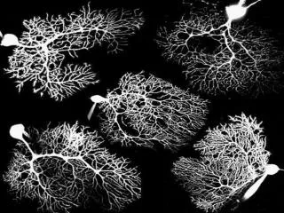
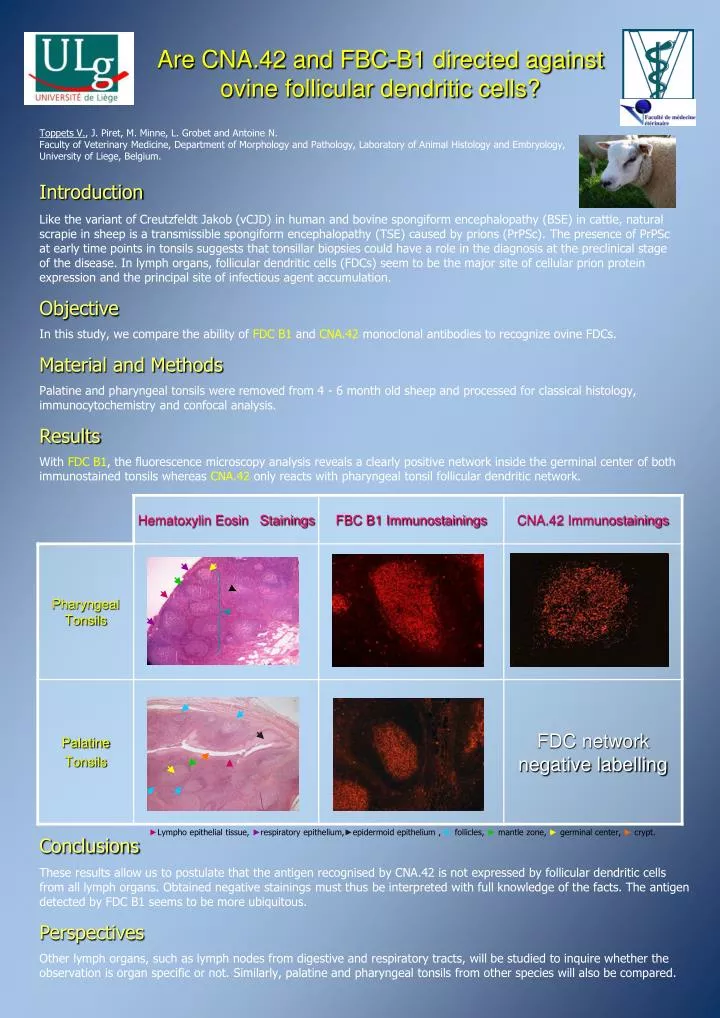
Toppets V., J. Piret, M. Minne, L. Grobet and Antoine N.Faculty of Veterinary Medicine, Department of Morphology and Pathology, Laboratory of Animal Histology and Embryology, University of Liege, Belgium. Are CNA.42 and FBC-B1 directed against ovine follicular dendritic cells? Introduction Like the variant of Creutzfeldt Jakob (vCJD) in human and bovine spongiform encephalopathy (BSE) in cattle, natural scrapie in sheep is a transmissible spongiform encephalopathy (TSE) caused by prions (PrPSc).The presence of PrPSc at early time points in tonsils suggests that tonsillar biopsies could have a role in the diagnosis at the preclinical stage of the disease. In lymph organs, follicular dendritic cells (FDCs) seem to be the major site of cellular prion protein expression and the principal site of infectious agent accumulation. Objective In this study, we compare the ability ofFDC B1and CNA.42monoclonal antibodies to recognize ovine FDCs. Material and Methods Palatine and pharyngeal tonsils were removed from 4 - 6 month old sheep and processed for classical histology, immunocytochemistry and confocal analysis. Results WithFDC B1, the fluorescence microscopy analysis reveals a clearly positive network inside the germinal center of both immunostained tonsils whereasCNA.42 only reacts with pharyngeal tonsil follicular dendritic network. ►Lympho epithelial tissue, ►respiratory epithelium,►epidermoid epithelium , ► follicles, ► mantle zone, ► germinal center, ► crypt. Conclusions These results allow us to postulate that the antigen recognised by CNA.42 is not expressed by follicular dendritic cells from all lymph organs. Obtained negative stainingsmust thus be interpreted with full knowledge of the facts. The antigen detected by FDC B1 seems to be more ubiquitous. Perspectives Other lymph organs, such as lymph nodes from digestive and respiratory tracts, will be studied to inquire whether the observation is organ specific or not. Similarly, palatine and pharyngeal tonsils from other species will also be compared.