Download
1 / 2
20 likes | 102 Views

E N D
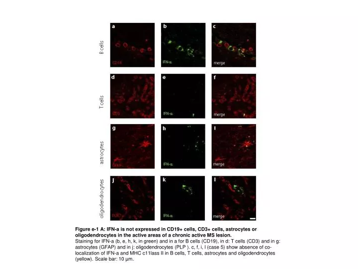
Figure e-1 A: IFN-a is not expressed in CD19+ cells, CD3+ cells, astrocytes or oligodendrocytes in the active areas of a chronic active MS lesion. Staining for IFN-a (b, e, h, k, in green) and in a for B cells (CD19), in d: T cells (CD3) and in g: astrocytes (GFAP) and in j: oligodendrocytes (PLP ). c, f, i, l (case 5) show absence of co-localization of IFN-a and MHC c11lass II in B cells, T cells, astrocytes and oligodendrocytes (yellow). Scale bar: 10 µm.
Figure e-1B: Plasmacytoid dendritic cells are not present in MS lesions. a-c: Immunohistochemical staining of formalin-fixed sections from MS autoptic specimens using plasmacytoid dendritic cell (pDC) markers as indicated. d-f: Immunohistochemical staining of formalin-fixed sections from a reactive lymph node using pDC markers as indicated. Note that CD123 also stains high endothelial venules. Scale bar: 200 µm. g: Immunohistochemical staining of BDCA2 in formalin-fixed normal brain. Scale bar: 100 µm.