Download
1 / 29
290 likes | 574 Views
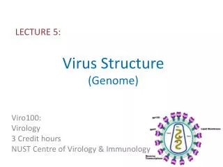
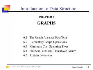
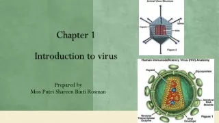
Introduction to Virus Structure. Tutorial Jonathan King, Peter Weigele, Greg Pintilie, David Gossard (MIT) v.November, 2008. Virus Structure. Size 17 nm – 3000 nm diameter Basic shape Rod-like “Spherical” Protective Shell - Capsid Made of many identical protein subunits

E N D
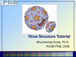
Introduction to Virus Structure Tutorial Jonathan King, Peter Weigele, Greg Pintilie, David Gossard (MIT) v.November, 2008
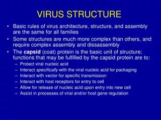
Virus Structure • Size • 17 nm – 3000 nm diameter • Basic shape • Rod-like • “Spherical” • Protective Shell - Capsid • Made of many identical protein subunits • Symmetrically organized • 50% of weight • Enveloped or non-enveloped • Genomic material • DNA or RNA • Single- or double-stranded
Virus Structure • Virus capsids function in: • Packaging and protecting nucleic acid • Host cell recognition • Protein on coat or envelope “feels” or “recognizes” host cell receptors • Genomic material delivery • Enveloped: cell fusion event • Non-enveloped: more complex strategies & specialized structures
Electron Microscopy Mitra, K. & Frank, J., 2006. Ribosome dynamics: insights from atomic structure modeling into cryo-electron microscopy maps. Annual review of biophysics and biomolecular structure, 35, 299-317.
History • In 1953, Crick & Watson proposed … principles of virus structure • Key insight: • Limited volume of virion capsid => nucleic acid sufficient to code for only a few sorts of proteins of limited size • Conclusion: • Identical subunits in identical environments • Icosahedral, dodecahedral symmetry
X-ray Crystallography of Viruses • Symmetry of protein shells makes them uniquely well-suited to crystallographic methods • Viruses are the largest assemblies of biological macromolecules whose structures have been determined at high resolution
History con’t • In 50’s & 60’s Klug and others confirmed that several (unrelated) “spherical” viruses had icosahedral symmetry • (Used negative staining & electron microscopy) • Conclusion: • Icosahedral symmetry is preferred in virus structure
Icosahedral Symmetry • 12 vertices • 20 faces (equilateral triangles) • 5-3-2 symmetry axes • 60 identical* subunits in identical environments can form icosahedral shell * asymmetric
But … • Clear evolutionary pressure to make larger capsid • Using larger subunits helps very little • Using more subunits helps a lot • Not possible to form icosahedral shell (of identical units in identical environments) with more than 60 subunits • Viruses with more than 60 subunits were observed • Question: • How can >60 subunits form an icosahedral shell? • Will any number of subunits work? • If so, how would they be organized?
Quasi-equivalence • In 1962, Caspar & Klug proposed the theory of “quasi-equivalence” • Not all protein subunits are equivalent • “Identical” subunits in slightly different environments • Only certain numbers of subunits will can be packed into closed regular lattice. Caspar & Klug, Cold Spring Harbor, 1962
Quasi-equivalence • Subunits are in “minimally” different environments • Pentamers at vertices • Hexamers elsewhere • Predicts packing arrangements of larger capsids • Shift from T1 to T4 packing => 8-fold increase in volume
HK97 Asymmetric Unit Outside Inside
Herpes Simplex Virus at 8.5 Å resolution from Wah Chiu and Frazer Rixon in Virus Research (2002)
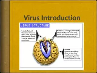
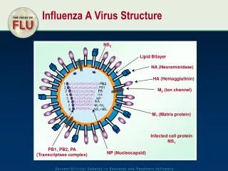
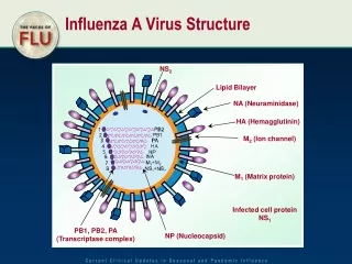
Influenza • Infection depends on spike proteins projecting from capsid membrane called “Hemagglutinin (HA)” • These bind sugar molecules on cell surface • Much of the difference between Hong Kong flu, Swine flu, Bird flu, and other strains, is in the amino acid sequence and conformation of the HA protein. • These differences control what host cell types the virus can infect. • Immunization against flu involves your immune system synthesizing antibody proteins that bind the HA protein.
Influenza virus composition of virus entry of influenza into cell
Influenza hemagglutinin: a pH induced, conformationally controlled trigger for membrane fusion 100 Å displacement of fusion peptide disordered loop backbone is structured low pH fusion peptide Qiao et al. Membrane Fusion Activity of Influenza Hemagglutinin. The Journal of Cell Biology, Volume 141, 1998
Influenza Hemagglutinin • The HA spikes extend like a spring during infection http://www.roche.com/pages/facets/10/viruse.htm http://hsc.virginia.edu/medicine/basic-sci/cellbio/jgruenke.html
Trimer Structure • Long alpha helices form coiled coil structure • In mature trimers of HA0, each monomer is cleaved into HA1 and HA2.
Evolution of dsDNA viruses • All known viruses, whether infecting bacteria or humans, may have evolved from a single common ancestor, relatively early in the evolution of organisms.
Common steps in the assembly of all dsDNA viruses • Unique portal ring at one Vertex • Scaffolding proteins • Procapsid assembled empty of DNA • DNA pumped into procapsid through portal ring • DNA moves back through portal to enter cell
Herpes viruses also have a portal protein portal complex Herpes portal (UL6) tagged with gold-bead labeled antibodies visualized by negative stain electron microscopy Bill Newcomb and Jay Brown, University of Virginia
Cryo-EM structure of purified Herpes portal protein Trus BL, Cheng N, Newcomb WW, Homa FL, Brown JC, Steven AC. Structure and polymorphism of the UL6 portal protein of herpes simplex virus type 1. J Virol. 2004 Nov;78(22):12668-71.