Download

1 / 31
360 likes | 602 Views
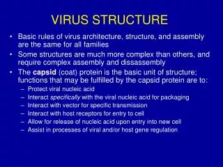
Virus Structure. Chapter 3. Tools for Studying Viral Structure. Electron Microscopy Excellent tool with some limitations High resolution Image can be a distortion due to specimen processing X-ray Diffraction Good for naked virions (no envelope) Cryoelectron Microscopy

E N D
Virus Structure Chapter 3
Tools for Studying Viral Structure • Electron Microscopy • Excellent tool with some limitations • High resolution • Image can be a distortion due to specimen processing • X-ray Diffraction • Good for naked virions (no envelope) • Cryoelectron Microscopy • Flash frozen with liquid nitrogen
The “virion” • Remember what it is? • Also known as a viral particle • Outside the host cell the virus exists as a virion. • Role of the virion: • Protects viral genome • Helps it gain entry to host cell
Structure of a Virus • Questions Relating to Structure • Is it rigid? • How big is it? • Is it flexible • Structure Must Serve Virus • It should provide protection for genome • It should allow virus to move from one host to next • It should allow for attachment of virus on to new host
Most fungal viruses have dsRNA genomes, • Most plant viruses have ssRNA genomes and • Most prokaryotic viruses have dsDNA genomes
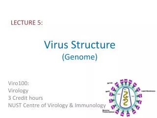
Viral Genome • Primary structure – encodes proteins required by virus • Secondary and tertiary structures also found as a way the virus controls gene expression. • pseudoknots: enzyme activity, ribosomal frameshifting • Internal ribosome entry sit (IRES)
Modifications at the ends of virusgenomes • covalently linked protein at the 5 end. In at least some viruses this is a vestige of a primer that was used for initiation of genome synthesis • Some genome RNAs have one or both of the modifications that occur in eukaryotic messenger RNAs (mRNAs): a methylated nucleotide cap at the 5 end and a sequence of adenosine residues (a polyadenylate tail; poly(A) tail) at the 3 end • The genomes of some ssRNA plant viruses are base paired and folded near their 3 ends to form structures similar to transfer RNA. These structures contain sequences that promote the initiation of RNA synthesis
Proteins non-covalently associatedwith virus genomes • Many nucleic acids packaged in virions have proteins bound to them non-covalently. • Rich in the basic amino acids lysine, arginine and histidine, which are –ve able to bind strongly to the +ve charged nucleic acids. • Papillomaviruses and polyomaviruses, (DNA viruses), have cell histones bound to the virus genome. • Most proteins associated with virus genomes, however, are virus coded, e.g HIV-1 nucleocapsid protein that coats the virus RNA; 29 per cent of its amino acid residues are basic. • Nucleic-acid-binding proteins may have other characteristics, such as zinc fingers the HIV-1 nucleocapsid protein has two zinc fingers. • In some viruses, such as tobacco mosaic virus, the protein coating the genome constitutes the capsid of the virion.
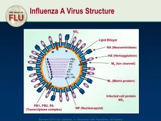
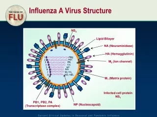
Segmented genomes • Genes of some viruses encoded in two or more nucleic acid molecules. • Segmented genomes are much more common amongst RNA viruses than DNA viruses Influenza A Virus
Repeat sequences • The genomes of many viruses contain sequences that are repeated. • These sequences include promoters, enhancers, origins of replication and other elements involved in virus replication. • Many linear virus genomes have repeat sequences at the ends (termini), in which case the sequences are known as terminal repeats • If the repeats are in the same orientation they are known as direct terminal repeats (DTRs), • Repeats in the opposite orientation they are known as inverted terminal repeats (ITRs).
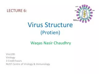
Virus proteins • Small genome viruses • The virion of tobacco mosaic virus contains only one protein species. • The virions of parvoviruses contain two to four protein species. These are viruses with • As the size of the genome increases, so the number of protein species tends to increase • Large genome viruses • The virion of herpes simplex virus 1 contains 39 protein species • The virion of the algal virus Paramecium bursaria Chlorella virus 1 contains over 100 protein species
Structural Proteins • Proteins that are components of virions are known as structural proteins. • Functions: • protection of the virus genome • attachment of the virion to a host cell (for many viruses) • fusion of the virion envelope to a cell membrane (for enveloped viruses)
Non-structural proteins • Proteins synthesized by the virus in an infected cell but they are not virion components). • Functions: • enzymes, e.g. protease, reverse transcriptase • transcription factors • primers for nucleic acid replication • interference with the immune response of the host.
Names of Viral Proteins • Structural proteins VP1, VP2, VP3, . . . (VP = virus protein) • Non-structural proteins: NSP1, NSP2, NSP3, . . .. • Many virus proteins are known by an abbreviation of one or two letters, which may indicate: • a structural characteristic G (glycoprotein) • P (phosphoprotein) • or a function F (fusion) • P (polymerase) • RT (reverse transcriptase).
Viral Capsid • Characteristics: • Protection – without capsid the genome is more susceptible to being inactivated. • Recognize and attach to host cell and then be able to release the genome. • A lipid envelope surrounds capsid in some viruses • An additional protein layer might also surround the capsid which is called the nucleocapsid.
Structural Symmetries • Icosahedral Symmetry • 20 triangular faces • It is a common capsid structure • Examples of viruses with icosahedral symmetry • Parvoviruses • These are simple viruses • 5 Kb ssDNA genome • Capsid is formed with 60 copies of single protein • Protein is approximately 520 a/a • 1/3 of genome is dedicated to capsid • Polio virus • Uses 180 copies of 3 subunit proteins • Much bigger virus
Viral Envelope • Lipid bilayer • Most originate from cellular host • Cholesterol and glycoproteins are present • In cases where budding occurs at the plasma membrane (Ex. Influenza) envelope resembles host’s plasma membrane i.e cholesterol and phospholipids • In cases where budding occurs at the ER (Ex. Flaviviruses) envelope has less cholesterol, similar to ER
Capsid • Helical • Icoshaderal
Icosahedral capsids a) Crystallographic structure of a simple icosahedral virus. b) The axes of symmetry
Helical Symmetry • The simplest way to arrange multiple, identical protein subunits is to use rotational symmetry & to arrange the irregularly shaped proteins around the circumference of a circle to form a disc. • Multiple discs can then be stacked on top of one another to form a cylinder, with the virus genome coated by the protein shell or contained in the hollow centre of the cylinder. • Tobacco mosaic virus (TMV) is representative of one of the two major structural classes seen in viruses of all types, those with helical symmetry.
Enveloped helical virus Enveloped icosahedral virus
Enveloped Structure of HIV Transmission Electron Micrograph of HIV-1 The nucleocapsid (arrows) can be seen within the envelope.
RNA viruses From Principles of Virology Flint et al ASM Press
DNA viruses From Principles of Virology Flint et al ASM Press
Viral Glycoproteins • Glycoproteins • Short cytoplasmic tail • Hydrophobic segment for anchoring (~20 amino acids) • Relatively large ectodomain (external domain) • Ectodomain • Extensively glycosylated preventing aggregation of virions • Glycosylation attracts water and reduces sticking (carried out in ER) • Palmitoylation of cysteine residues is also extensive (carried out in ER) • Most envelope proteins are type I • That means N-terminus facing out, C terminus near anchor domain • Some though are type II