Download
1 / 76
770 likes | 905 Views
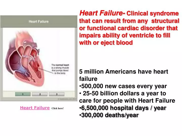
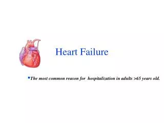
Heart Failure- Clinical syndrome that can result from any structural or functional cardiac disorder that impairs ability of ventricle to fill with or eject blood 5 million Americans have heart failure 500,000 new cases every year

E N D
Heart Failure- Clinical syndrome that can result from any structural or functional cardiac disorder that impairs ability of ventricle to fill with or eject blood • 5 million Americans have heart failure • 500,000 new cases every year • 25-50 billion dollars a year to care for people with Heart Failure • 6,500,000 hospital days / year • 300,000 deaths/year Heart Failure Click here!
Heart Failure Cardiogenic shock Cardiomyopathy Severe End Stage Mild Mild Pulmonary Edema Control With /Same as Mild with Morphine Sulfate Irreversible Needs new ventricle VAD IABP Drugs Diet Fluid Restriction VAD IABP Heart Transplant
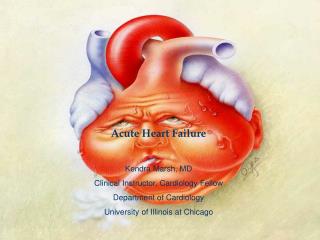
Definition-Heart Failure • CO = SV x HR is insufficient to meet the metabolic needs of the body • SV is determined by preload, afterload and myocardial contractility • Systolic failure- dec. contractility • Diastolic failure- dec. filling • EF< 40%
Heart Failure Pneumonic(common therapies) U Upright Position N Nitrates L Lasix O Oxygen A ACE, ARBs, Amiodorone D Dig, Dobutamine M Morphine Sulfate E Extremities Down
Heart Failure Click here for Online Lecture (Interactive) or Click here for Online Lecture (Read)
Keys • all organs (liver, lungs, legs, etc.) return blood to heart • . When heart begins to fail/ weaken- unable to pump blood forward-fluid backs up and increases pressure within all organs. • Organ response • LUNGS: congested-become “stiffer” , inc effort to breathe; fluid starts to escape into alveoli; fluid interferes with oxygen exchange, aggravates shortness of breath. • Shortness of breath during exertion, may be one of earliest symptoms; then progresses; later requiring extra pillows at night to breathe; the experience "P.N.D." or paroxysmal nocturnal dyspnea . • Pulmonary or lung edema • Legs, ankles, feet- blood from feet and legs; in failing heart, there is a back-up of fluid and pressure in these areas, heart is unable to pump blood as promptly as received; increased fluid within feet and legs causes fluid to "seep" out of blood vessels ; increased body
Factors effecting heart pump effectiveness Preload • Volume of blood in ventricles at end diastole • Depends on venous return • Depends on compliance Afterload • Force needed to eject blood into circulation • Arterial B/P, pulmonary artery pressure • Valvular disease increases afterload
Cardiomegaly/ventricular remodeling occurs as heart overworked-changes in size, shape, and function of the heart after injury to left ventricle. Injury due to acute myocardial infarction or due to causes that increase pressure or volume overload on the heart (Heart failure)
Heart Failure (AKA-congestive heart failure) • Pathophysiology • A. Cardiac compensatory mechanisms • 1.tachycardia • 2.ventricular dilation-Starling’s law • 3.myocardial hypertrophy • Hypoxia leads to dec. contractility
cont. • B.Homeostatic Compensatory mechanisms • Sympathetic Nervous System-(beta blockers block this) • 1. Vascular system- norepinephrine- vasoconstriction (What effect on afterload?) • 2. Kidneys- • A. Dec. CO and B/P cause renin angiotensin release. (ACE) • B. Aldosterone release causes Na and H2O retention • Inc. Na causes release of ADH (diuretics) • Release of atrial natriuretic factor- promotes Na and H20 excretion, prevents severe cardiac decompensation • 3. Liver- stores venous volume(ascites, +HJR, Hepatomegaly- can store 10 L. check enzymes
Result of Compensatory Mechanisms Heart Failure
Pathophysiology-Structural Changes with HF • Decreased contractility • Increased preload (volume) • Increased afterload (resistance) • Ventricular remodeling (ACE inhibitors can prevent this) • Ventricular hypertrophy • Ventricular dilation
END RESULT FLUID OVERLOAD AND Acute Decompensated Heart Failure (ADHF) or Pulmonary Edema
ACC/AHA Stages NY ASSN Funct Class
CHF-Causes • 1. Impaired cardiac function • Coronary heart disease • Cardiomyopathies • Rheumatic fever • Endocarditis • 2. Increased cardiac workload • Hypertension • Valvular disorders • Anemias • Congenital heart defects • 3.Acute non-cardiac conditions • Volume overload • Hyperthyroid, Fever,infection
Classifications • Systolic versus diastolic • Systolic- loss of contractility get dec. CO • Diastolic- decreased filling or preload • Left-sided versus right –sided • Left- lungs • Right-peripheral • High output- hypermetabolic state • Acute versus chronic • Acute- MI • Chronic-cardiomyopathy
Left Ventricular Failure • Signs and symptoms • dyspnea • orthopnea PND • Cheyne Stokes • fatigue • Anxiety • rales • NOTE L FOR LEFT AND L FOR LUNGS
Pulmonary Edema(advanced L side HF) • When PA WEDGE pressure is approx 30mmHg • Signs and symptoms • 1.wheezing • 2.pallor, cyanosis • 3.Inc. HR and BP • 4.s3 gallopThe Auscultation Assistant - Rubs and Gallops • 5.rales,copious pink, frothy sputum
Person literally drowning in secretions Immediate Action Needed
Acute Decompensated Heart Failure (ADHF) Pulmonary Edema As the intracapillary pressure increases, normally impermeable (tight) junctions between the alveolar cells open, permitting alveolar flooding to occur. Pulmonary edema begins with an increased filtration through the loose junctions of the pulmonary capillaries.
Goals of Treatment • MAD DOG • Improve gas exchange • O2 • intubate • elevate HOB
Right Heart Failure • Signs and Symptoms • fatigue, weakness, lethargy • wt. gain, inc. abd. girth, anorexia,RUQ pain • elevated neck veins • Hepatomegaly +HJR • may not see signs of LVF
Can Have RVF Without LVF • What is this called? COR PULMONALE
Diagnostic Tests • CXR • EKG and cardiac enzymes • Electrolytes, BUN and Creat • Liver function tests • Hemodynamic Monitoring-CVP and SG • Echo to determine EF%(ejection fraction) • May be stressed with exercise or medicine • Cardiac Cath to determine heart pressures ( inc.PAW • Signs and symptoms of HF • *BNP(beta natriuretic peptide) 0-100
Nursing Assessment • Vital signs • PA readings • Urine output • -What else!!
Decreased cardiac output • Plan frequent rest periods • Monitor VS and O2 sat at rest and during activity • Take apical pulse • Review lab results and hemodynamic monitoring results • Fluid restriction- keep accurate I and O • Elevate legs when sitting • Teach relaxation and ROM exercises
Activity Intolerance • Provide O2 as needed • practice deep breathing exercises • teach energy saving techniques • prevent interruptions at night • monitor progression of activity • offer 4-6 meals a day
Fluid Volume Excess • Give diuretics and provide BSC • Teach side effects of meds • Teach fluid restriction • Teach low sodium diet • Monitor I and O and daily weights • Position in semi or high fowlers • Listen to BS frequently
Knowledge deficit • Low Na diet • Fluid restriction • Daily weight • When to call Dr. • Medications
Impaired Skin Integrity • YOU ALL KNOW THIS ONE
Ineffective Breathing PatternImpaired gas Exchange • Observe for signs of resp distress • Monitor O2 sats and ABGs • What else
GOALS • Decrease preload • Dec. intravascular volume • Dec venous return • Fowlers • MSO4 and Ntg • Decrease afterload • Inc. cardiac performance(contractility) • CRT • Balance supply and demand of oxygen • Inc. O2- O2, intubate, HOB up,Legs down, mech vent with PEEP • Dec. demand-beta blockers, rest, dec B/P Manage symptoms
CRT-Cardiac Resynchronization Therapy HOW IT WORKS: Standard implanted pacemakers are equipped with two wires (or "leads") that conduct pacing signals to specific regions of the heart (usually at positions A and C). The biventricular pacing devices have added a third lead (to position B) that is designed to conduct signals directly into the left ventricle. The combination of all three leads creates a synchronized pumping of the ventricles, increasing the efficiency of each beat and pumping more blood on the whole.

![Heart Failure [HF]](https://cdn2.slideserve.com/5103874/heart-failure-hf-dt.jpg)