Download
1 / 10
100 likes | 258 Views
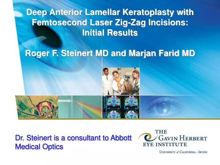
Deep Anterior Lamellar Keratoplasty with Femtosecond Laser Zig-Zag Incisions: Initial Results Roger F. Steinert MD and Marjan Farid MD. Dr. Steinert is a consultant to Abbott Medical Optics. Purpose.

E N D
Deep Anterior Lamellar Keratoplasty with Femtosecond Laser Zig-Zag Incisions: Initial ResultsRoger F. Steinert MD and Marjan Farid MD Dr. Steinert is a consultant to Abbott Medical Optics
Purpose • To evaluate visual recovery and astigmatism in eyes with stromal corneal pathology and ecstatic disease that underwent deep anterior lamellar keratoplasty (DALK) with femtosecond laser zigzag incisions
Methods • Retrospective Chart Review • Patient selection: • Consecutive patients who underwent femtosecond laser assisted DALK over a 1 year period • Exclusion: Cystic blebs, severe peripheral opacity or NV
Main Outcome Measures • Astigmatism (by Orbscan or Pentacam) • BSCVA
Surgical Technique 1 • Femtosecond laser zig-zag incision:The femtosecond laser is used to create matching zig-zag (posterior angled cut, lamellar ring cut at 300um depth and anterior angled cut) incisions in the host and donor corneas. Based on the thinnest area of the patient’s peripheral cornea, the laser is set to leave 70 microns of posterior uncut tissue. • DALK:The zig-zag incision is bluntly dissected. A 30-gauge needle on an air-filled syringe is inserted along the lamellar ring cut or just above the 70 micron uncut tissue. Air is injected at this level to bare Descemet’s membrane with the big-bubble technique as described by Anwar.
Surgical Technique 2 • If successful, the remainder of the posterior stromal cut is excised, leaving bare Descemet’s membrane. The endothelium is wiped off the donor cornea and the donor is sutured to the patient’s corneal rim. • If a Descemet’s membrane perforation occurs, the surgeon converts to a standard femtosecond laser assisted PKP. In that case, the donor corneal endothelium is left intact before suturing it to the corneal rim.
Results • Average BSCVA by month 3 was 20/32 (logMar 0.2) • In 4 out of 6 patients who had topography performed at that visit: • Average astigmatism by month 1 was 2.45 diopters (1.26 SD) • Average astigmatism by month 3 was 2.7 diopters (1.22 SD)
Results • Patient 4 had recurring epithelial breakdown due to HSV keratitis • Approximately 30% of our initial big bubble attempts resulted in Descemet’s perforations which required conversion to full thickness PKP • Visante OCT showed good Descemet’s membrane apposition to the donor cornea and the zig-zag wound healing reaction in all eyescses.
Conclusions • Femtosecond zigzag DALK results in a biomechanically stable wound and excellent apposition of Descemet’s membrane. • Early results demonstrate excellent recovery of BSCVA and astigmatism which are comparable to full thickness femtosecond laser keratoplasty. • There is an early surgical learning curve for achieving successful baring of Descemet’s membrane