Download
1 / 37
390 likes | 663 Views
Lipid breakdown and biosynthesis. Chemistry 256. Lipids are tied to metabolism through the TCA cycle.

E N D
Lipid breakdown and biosynthesis Chemistry 256
Lipids are tied to metabolism through the TCA cycle The dihydroxyacetone phosphate (DHAP) made by glycolysis or made from oxaloacetate by glyceroneogenesis; thus this system responds to the same hormones involved in regulation of carbohydrate metabolism.
Lipids are degraded by digestion prior to absorption • Triacylglycerols are 90% of dietary lipids. • Digestion of these occurs at the lipid-water interface since lipids are insoluble in water and digestive proteins are soluble. • Bile acids (or bile salts) are cholesterol derivatives that help emulsify the mixture in the small intestine. • Lipases are the class of proteins that digest the triacylglycerols.
Lipid digestion by lipases occurs by a variety of mechanisms All lipases convert triacylglycerols into fatty acids, monoacylglycerols or diacylglycerols. The specific mechanisms vary; phospholipase A2 has a hydrophobic channel that allows lipids to enter active site.
Lipid droplets are broken up into micelles (or at least smaller membranes) which are then broken up into fatty acids and other smaller parts which are absorbed by the intestinal cells.
Further in the cell, the fatty acids end up in the ER and are packaged into chylomicrons (a type of lipoprotein), which are then extruded into the lymphatic system.
VLDLs, IDLs and LDLs are synthesized by the liver for lipid transport to tissues; tissues synthesize HDLs for transport to liver. HDLs are the smallest particles and the most rich in triacylglycerols.
Apolipoproteins surround the lipoproteins to hold them together. Apolipoprotein B-100 (apoB-100) is a big (4536-residue) monomeric protein with distinct polar and nonpolar moieties to interact with membranes and the lipoprotein. Note the presence of cholesteryl ester in the lipoprotein; it is a source of cholesterol for cells.
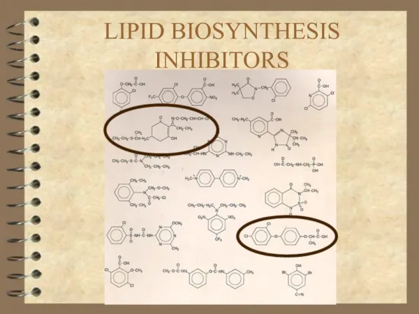
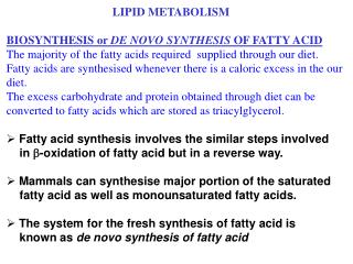
Breakdown versus synthesis • Major difference: Even though two-carbon units are added to the growing fatty acid chain, it is malonyl-CoA (3 carbons), not acetyl-CoA (2 carbons) that is added. • In addition, bicarbonate is a required substrate. • Of course, this being a reduction, a lot of energy-bearing molecules will be oxidized. • So: many different enzymes than β-oxidation!
The sites of these activities • In much the same way that hormones like glucagon and insulin regulate the glycolytic and glucogenolytic pathways, these hormones regulate the breakdown and buildup of fatty acids. • Hormone-sensitive triacylglycerol lipase is turned on and off via phosphorylation and dephosphorylation, which is triggered by cAMP concentration.
Fatty acid oxidation Depending on the original number of carbons, the excretion product of fatty acid oxidation varies. This experiment by Franz Knoop in 1904 (University of Freiburg) used benzene-derivatized fatty acids fed to dogs, and showed that the fatty acids were degraded two carbons at a time.
Fatty acids are activated by their attachment to coenzyme A, facilitated by acyl-CoA synthetases. These are located on the ER or the outer mitochondrial membrane. Mixed anhydride intermediate initiated by a nucleophilic attack on ATP α-phosphorus.
Fatty acids are oxidized in the mitochondrial matrix, and thus must be transported in. The acyl portion of the fatty acid is transported and reattached to CoA inside the matrix.
Fatty acid oxidation to produce a single acetyl-CoA unit also produces a FADH2,which is oxidized by the standard electron-transport chain, for a further 1.5 ATP. Note the formation of a trans-fatty acid in the first step.
The fourth step (the one that produces the acetyl-CoA) involves a thiolase, which proceeds as a Claisen ester cleavage (the reverse of a Claisen condensation). Much energy can thus be produced from a single fatty acid; one palmitate (C16) yields 106 ATP.
Unsaturated fatty acids require an additional set of enzymes, because an adjacent set of double bonds cannot be formed. Note the usage of some energy to achieve this. Odd-numbered fatty acids yields propionyl-CoA, which is converted to succinyl-CoA to enter the TCA.
Dorothy Hodgkin in 1956 (Oxford; Nobel Prize, 1964) determined the structure of the enzyme involved in producing succinyl-CoA, using X-ray crystallography. It is a rare cobalt-containing enzyme; note its position in a heme-like ring.
Ketone bodies are produced by the liver mitochondria as an alternate fuel source for tissues like the heart and other muscles, or the brain during starvation.
Fatty acid biosynthesis begins with acetyl-CoA The malonyl-CoA then reacts with an acyl-carrier protein (ACP) through a thiol group to make malonyl-ACP; an acetyl-CoA does the same reaction to produce acetyl-ACP. The malonyl-ACP and acetyl-ACP react to make acetoacetyl-ACP, with the loss of a CO2 and an ACP.
Much energy is required: To make palmitic acid, in addition to 8 acetyl-CoA, 14 NADPH and 7 ATP are required. The enzyme complex is fatty acid sythase, which is (not surprisingly) a multi-function enzyme. Other enzymes are required to form unsaturations.
Citrate shuttle allows acetyl-CoA out of mitochondrion The site of fatty acid (and other lipid) biosynthesis is in the cytosol (usually the endoplasmic reticulum), but the acetyl-CoA precursor is found in the mitochondria. The inner mitochondrial membrane, as might be expected, is impermeable to acetyl-CoA, so the acetyl-CoA is converted to pyruvate and then to citrate and transported out of the mitochondrion, and then reconverted to acetyl-CoA (similar to the aspartate/malate shuttle for gluconeogenesis).
Acetyl-CoA carboxylase produces malonyl-CoA ACC is a 230-kD polypeptide (shown below), first step in turning acetyl-CoA into a fatty acid. Citrate is an activator; fatty acids are an inhibitor (feedback). Serine 79 is a phosphorylation site; phosphorylation by AMP-dependent protein kinase inactivates enzyme. Glucagon and epinephrine activate protein kinase A, which inhibits dephosphorylation of serine 79, and thus inhibits ACC; insulin promotes dephosphorylation, which activates ACC. α-ACC in adipose tissue; β-ACC in heart tissue
Triacylglyceride synthesis Independent of fatty acid synthesis, triacylglycerol synthesis begins with a glycolysis intermediate, dihydroxyacetone phosphate (DHAP) undergoing reduction by glycerol-3-P dehydrogenase to glycerol-3-phosphate. Interestingly, the loss of glycerol-3-phosphate by liver tumor cells is part of the mechanism of cancer (Howard, Morris and Bailey, “Ether-lipids, a-glycerol phosphate dehydrogenase, and growth rate in tumors and cultured cells”, Cancer Res., 1972)
Triacylglyceride synthesis The fatty acid comes in, now, as the R (acyl) group added with the help of glycerol-3-phosphate acyltransferase (first step here).
Triacylglyceride synthesis The previous slide mechanism is found in the endoplasmic reticulum and mitochondrion; this alternate pathway exists in peroxisomes and bacteria, in which the acyl transfer takes place without going through glycerol-3-phosphate (Schjuman and de Mendoza, “Solving an old puzzle in phospholipid biosynthesis”, Nature Chemical Biology 2006) In any case, both pathways end at the same molecule: lysophosphatidic acid, and then on to triacylglycerides as the other two acyl groups are transferred…however, even as the second fatty acid group is transferred, other types of molecules can be made.
The synthesis of glycerophospholipids • Note in this mechanism, the head group is phosphorylated and then the diacylglycerol is transferred. • In the second step, cytidine triphosphate (CTP – a nucleotide) is used as an energy source and a phosphate source.
The synthesis of sphingolipids • Sphingolipids deviate from the triacylglyceride synthesis path; instead, these molecules start from t palmitoyl-CoA and another acyl-CoA, as well as the amino acid serine. • At this point, the head group is modified to include carbohydrate units (ceramides).
The initiation of eicosanoid lipids • To activate the arachidonic acid, an unusual peroxide structure is formed, requiring prostaglandin H2 synthase which has the cyclooxygenase and peroxidase function. • Aspirin’s mechanism involves acetylating a serine residue of prostaglandin H2 synthase which prevents the formation of PGH2, a precursor to the pain signal prostaglandins (Vain, “Inhibition of prostaglandin synthesis as a mechanism of action for aspirin-like drugs”, Nature New Biology, 1971).
Steroid synthesis - the first steps (the amino acid residue numbers refer to a HMG-CoAsynthase enzyme)
Steroid synthesis • HMG-CoA ( = 3-hydroxy-3-methylglutaryl-coenzyme A) is a necessary precursor. Note the HMG unit is modified into the isoprenoid structure on this molecule.
Steroid synthesis • The C30 cholesterol precursor squalene is built from the condensation of six isoprene units, though a farensyl intermediate. • The farensyl intermediate is made through the head-to-tail condensation of three isoprenoid units, then the squalene is made through the head-to-head condensation of two farensyl units.
Steroid synthesis • Another unusual structure, an epoxide (catalyzed by squalene epoxidase) leads to the placement of the hydroxy group at C-3.
Steroid synthesis • The cyclization that results in the fused ring structure of cholesterol begins as a consequence of the reduction of the epoxide at C-3 by oxidosqualene cyclase.
Cholesterol synthesis • The modification of lanosterol to cholesterol is a 19-step process and occurs in the ER membrane. In the liver, the C-3 alcohol can be esterified to make a cholesteryl ester.