Download
1 / 17
170 likes | 363 Views
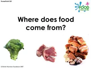
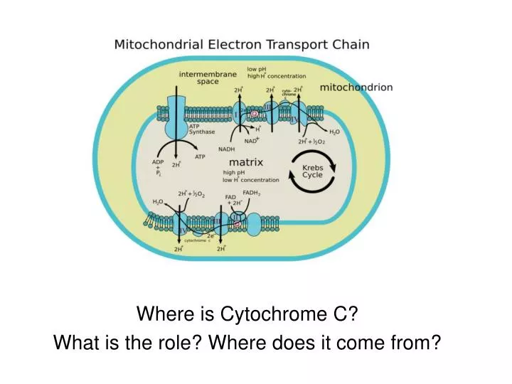
Where is Cytochrome C? What is the role? Where does it come from?. Evolutionary Biology of Cytochrome C. Cytochrome c is a highly conserved protein. Its primary structure consists of a chain of 100 aa.

E N D
Where is Cytochrome C? What is the role? Where does it come from?
Evolutionary Biology of Cytochrome C • Cytochrome c is a highly conserved protein. • Its primary structure consists of a chain of 100 aa. • Both chicken and turkeys have the identical molecule whereas ducks p differ by one amino acid. • Similarly, both humans and chimpanzees have the identical molecule.
Where does protein come from? The information for building proteins is stored in our DNA. DNA codes for proteins like cytochrome C. DNA RNA Protein Cytochrome C Gene for Cytochrome C Gene expression If the DNA in a single human cell were stretched out and laid end-to-end, it would measure approximately 6.5 feet. The average human body contains 10 to 20 billion miles of DNA distributed among trillions of cells.
Understanding How Cells Build Essential Proteins DNA Structure Double helix WC Base pair Structure of a Gene Understanding how genes are expressed DNA RNAProtein
Twist DNA is a double-stranded helix James Watson and Francis Crick Worked out the three-dimensional structure of DNA, based on work by Rosalind Franklin http://www.dnai.org/ Figure 10.3A, B
Sugar-phosphate backbone Phosphate group Nitrogenous base A A Sugar Nitrogenous base(A, G, C, or T) Phosphategroup C C DNA nucleotide O H H3C C C N O C C T CH2 H T O P N O O O– Thymine (T) O C C H H H H G G C C H O Sugar(deoxyribose) T T DNA nucleotide DNA polynucleotide DNA is a nucleic acid • Made of long chains of nucleotide monomers • Remember that ATP was a nucleotide Figure 10.2A
Double Helix StructureMajor/minor grooveAnti-parallel strands
H H H H O N N O C H C H H3C C C H N N N C C N C N N C H H C C C C C C C C C C H O H N N N O H N N H N N H H H H H Adenine (A) Guanine (G) Thymine (T) Cytosine (C) Purines Pyrimidines DNA has four kinds of nitrogenous bases • A, T, C, and G Figure 10.2B
5 end 3 end P HO 5 4 2 3 A T 3 1 1 4 2 5 P P C G P P G C P P A T OH P 3 end 5 end Each strand of the double helix is oriented in the opposite direction Figure 10.5B
Understanding How Cells Build Essential Proteins DNA Structure Double helix WC Base pair Structure of a Gene Understanding how genes are expressed DNA RNAProtein DNA nevers leaves the nucleas but the information does!
Strand to be transcribed T A C T T C A A A A T C DNA A T G A A G T T T T A G Transcription G U U U A G A U A A G U RNA Startcondon Stopcondon Translation Met Polypeptide Lys Phe Figure 10.8B • The information constituting an organism’s genotype • is carried in its sequence of its DNA bases. • “ATGACTAA” • A particular gene, a linear sequence of many nucleotides • Specifies a polypeptide • “MET-LYS-LEU”
DNA Transcription RNA Translation Protein Figure 10.6A
RNA nucleotides RNA polymerase A C C A T T A U T C T G U G A C A U C C A C C A G A T T T A G G Direction of transcription Template Strand of DNA Figure 10.9A Newly made RNA DNA RNA Protein Close up on DNARNA: Transcription
RNA nucleotides RNA polymerase A C C A T T A U C T T G U G A C A U C C A C C A G A T T T A G G Direction of transcription Template Strand of DNA Figure 10.9A Newly made RNA DNA RNA Protein transcription • In the nucleus, the DNA helix unzips • And RNA nucleotides line up along one strand of the DNA, following the base pairing rules • As the single-stranded messenger RNA (mRNA) peels away from the gene • The DNA strands rejoin
RNA polymerase DNA of gene Promoter DNA Terminator DNA Area shown In Figure 10.9A Growing RNA Completed RNA RNA polymerase Figure 10.9B DNA RNA Protein • RNA pol binds to promoter • Transcription of strand to mRNA • Instructions now in mRNA • RNA pol reaches terminator
DNA molecule Gene 1 Gene 2 Second base U C A G U UAU UGU UGC UGA Stop UUU UCU Gene 3 Cys Phe Tyr UUC UAC C UCC Ser U UCA UUA UAA Stop A Leu UCG UAG Stop UGG Trp G U CAU CGU CUU CCU His DNA strand C CAC CGC CUC CCC C Pro Arg Leu A A A C A C G G A A C A CUA CCA CAA CGA A Gln Third base CAG CGG CUG CCG G First base Transcription U ACU AUU AAU AGU Ser Asn ACC AGC AUC AAC Ile C U U U G U G C C U U G U A Thr RNA AUA AGA ACA AAA A Lys Arg Met or start ACC AGG AAG AUG G Codon U GUU GAU GGU GCU Translation Asp C GGC GCC GUC GAC Gly Ala G Val GUA GCA GGA GAA A Glu GUG GCG GGG GAG Polypeptide G Figure 10.8A Amino acid Figure 10.7 DNA RNA Protein