Download
1 / 8
80 likes | 296 Views
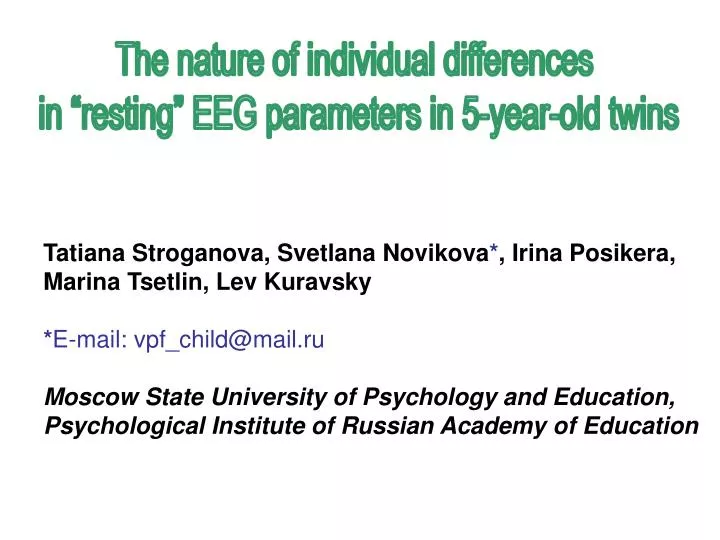
The nature of individual differences in “resting” EEG parameters in 5-year-old twins. Tatiana Stroganova, Svetlana Novikova * , Irina Posikera, Marina Tsetlin, Lev Kuravsky * E-mail: vpf_child@mail.ru Moscow State University of Psychology and Education ,

E N D
The nature of individual differences in “resting” EEG parameters in 5-year-old twins Tatiana Stroganova, Svetlana Novikova*, Irina Posikera, Marina Tsetlin, Lev Kuravsky *E-mail: vpf_child@mail.ru Moscow State University of Psychology and Education, PsychologicalInstitute of Russian Academy ofEducation
Introduction Background EEG measures have been repeatedly reported to depend exclusively on genetic influences in adults and adolescents (Lykken et al., 1982;van Beijsterveldt et al., 1996), but there have been few genetic studies of smaller children. There are just one study devoted to EEG genetics in infants (Orekhova et al., 2003) and one devoted to preschoolers (van Baal et al., 1996). Under various experimental conditions, the same EEG parameters may reflect different brain processes. Therefore, the contribution of genetic and environmental factors may depend on the current functional state. Little is known about the sources of individual differences of EEG parameters measured under experimental conditions other than the so-called baseline (quiet wakefulness with eyes closed). We estimated relative contribution of genetic and environmental factors to alpha gravity frequency and spectral amplitudes of three frequency bands in preschool EEG under two similar test conditions.
Methods Participants: 21 pairs of MZ and 20 pairs of same-sex DZ twins aged 5-6 years. All participants were bornwithin 32 to 41weeks of gestational age (mean=37.2 weeks,SD=1.91) and had no any known medical problems. Zygosity wasdetermined according to the Goldsmith questionnaire (Goldsmith, 1991). Procedure: EEG was registered during two “resting” conditions of dark homogeneous visual field, closed eyes and darkness, and one control condition of visual attention to moving color stimuli. On average, 40 s of the artifact-free EEG record for each condition were analyzed for each subject. Recording and processing of EEG:12 electrodes were placed at AF3, AF4, C3, C4, F7, F8, T5, T6, P3, P4, O1, and O2 positions. Linked ears served as the reference. Epochs of 2.5 s length were Fast Fourier Transformed (80% overlap) to yield the amplitude spectrum between 1.2 and 15.2 Hz in discrete bins of 0.4 Hz. Age-adjusted (Orekhova et al, 2006) frequency boundaries of three EEG bands – delta (1.2-3.2 Hz), theta (3.6-7.6 Hz), and alpha (8.0-12.8 Hz) – were used. Alpha gravity frequency (Lykken et al., 1982) was measured at the occipital region in range of 7-11 Hz (estimated in 94% of cases). Statistical analysis: ANOVA was performed to assess the differences between two test conditions in alpha gravity frequensy and in reactivity of spectral amplitudes. Fisher intrapair twin correlation and model fitting methodology (confirmatory factor analysis)were used for genetic analysis. The concordance of results obtained for two similar functional loads improves reliability of estimation. So we report only results reproduced in two test conditions. Preliminary analysis of genetic data: Analysis of variance has shown no main effects of sex and zygosity or their interactions that were reproduced in two subsamples that included one twin of each pair.
Result 1. Reactivity of absolute amplitudes under two test conditions. Spectral amplitudes of alpha frequency band changed quite similar from control to each of test conditions demonstrating significant increase. The same result was found for theta band amplitudes. Reactivity of delta amplitudes (near 3 Hz) was reliably greater under closed eyes at posterior regions (see arrows). The two test conditions are not equated in amount of horizontal eye movements. Horizontal eye movements disappear when eyes are closed but remain under darkness condition producing additive stimulation of cortex. Therefore different delta reactivity could reflect different degree of partial cortex deafferentation in two test conditions (Steriade et al., 1990). Alpha gravity frequency was near 8.5 Hz and did not significantly differ between two test conditions. Figure 1. Average amplitude spectra under two experimental and one control conditions (N=75). Visual attention Closed eyes Darkness
Result 2. Assessing of intrapair twin resemblance (correlational analysis). Figure 2. Intraclass correlations in the MZ and the DZ pairs for common logarithms of absolute spectral amplitudes. А – closed eyes, B – darkness. Ovals indicate highly significant (** - p<0.01; *** - p<.001) correlations in MZ and DZ pairs over parietal regions within theta band in both test conditions. Concordance of results under both test conditions was obtained for alpha as for theta band. We suggest variability of alpha amplitudes to depend essentially on genetic, probably non-additive factors (all rMZ except anterior regions are highly signifi-cant: p<.001; all rDZ are ns). Remarkably, alpha frequency has most likely nonadditive heritability too (rMZ≈0.8; rDZ≈0.0). Variability of parietal theta amplitudes perhaps is influenced by shared environment (rMZ≈rDZ).
Result 3. Model fitting confirmed all findings of the correlational analysis. Figure 3. The percent of EEG spectral amplitude variance explained by black- heritability, gray - shared environment, and white- nonshared environment estimates derived from the ace model with 3 degrees of freedom.A – closed eyes, B – darkness.4anteriorregions were excluded because of artifacts. For spectral amlitudes within alpha and theta but not delta band, results of model fitting for darkness condition actually mirror results during closed eyes condition. The best fitting models for alpha amplitudes always included genetic (additive or nonadditive) component. Similarly, nonadditive genetic model (de) satisfactorily explained variance of alpha frequency. The best fitting model for theta amplitudes at leads P3, P4, and T5 was acemodelwith equated contribution ofgenetic (a) andshared environmental (c) factors (4 degrees of freedom).
Conclusion 1. The results suggest that the sources of individual differences for oscillations within alpha (8.0-12.8 Hz) as for theta (3.6-7.6 Hz) frequency band did not differ between two conditions of homogenous field of vision: darkness and closed eyes. The nature of individual differences in variability of delta (1.2-3.2 Hz) amplitudes remained unclear since poor reproducibility of delta results in two test conditions. 2. Variance of alpha frequency as for alphaamplitudes depended mainly on non-additivegenetic factors in agreement with previous adult’s (Lykken et al., 1982;Posthuma et al., 2001) and children’s results (Orekhova et al., 2003) though these distinct parameters apparently have different nature. 3. The influence of shared environment is probable for theta amplitude at associative (first of all parietal) regions. This finding contradicts Dutch study of preschoolers (van Baal et al., 1996) that showed strong heritability of theta amplitudes. At the same time our finding is in agreement with Plomin’s amplification theory and with results of recent studies, reported rather powerful environmental contribution in infant theta amplitudes (Orekhova et al., 2003) and in adolescent females’ ERP amplitude and low-frequency EEG activity (Anokhin et al., 2001) during the conditions of dark homogenous visual field. 4. Both test conditions are accompanied by theta increase. We suppose that the reduced visual sensory input is unpleasant, emotionally loaded situation for 5-year-olds. Theta rhythm is well known to synchronize during emotional load (see Orekhova et al., 2006, for references). Cortical theta is provided by increased activity of limbic system and related structures, so called theta response system (Demiralp et al., 1994). 5. Taking into account functional role of theta sinchronization we conclude that environmental experience can modify the activity of structuresinvolved in regulation of slightly stressful functional load (not resting state!) in preschoolers under conditions of reduced visual sensory input. This inference includes our study in wide context of contemporary research of placticity of cortico-lymbic structures in early human and animal development (Chugani et al., 2001;Plotsky & Meaney, 1993).
Anokhin A.P., van Baal G.C.M., van Beijsterveldt C.E.M., de Geus E.J.C., Grant G., Boomsma D.I. (2001). Genetic correlation between the P300 event-related brain potential and the EEG power spectrum.Behavior Genetics, 31. 545-554. Chugani H.T., Behen M.E., Muzik O., Juhász C., Nagy F., Chugani D.C. (2001). Local brain functional activity following early social deprivation: A study of postinstitutionalized Romanian orphans. NeuroImage, 14, 1290–1301. Demiralp T., Başar-Eroglu C., Rahn E., Başar E. (1994). Event-related theta rhythms in cat hippocampus and prefrontal cortex during an omitted stimulus paradigm. Int J Psychophysiol., 18. 35-48. Goldsmith HH. (1991). A zygosity questionnaire for young twins: A research note. Behavior Genetics, 21. 257–269. Lykken D., Tellegen A., Iacono W.G. (1982). EEG spectra in twins: evidence for a neglected mechanism of genetic determination. Physiological psychology, 10. 60-65. Orekhova E.V., Stroganova T.A., Posikera I.N., Malykh S.B. (2003). Heritability and “environmentability” of electroencephalogram in infants: The twin study. Psychophysiology, 40. 727-741. Orekhova E.V., Stroganova T.A., Posikera I.N., Elam M. (2006). EEG theta rhythm in infants and preschool children. Clin Neurophy- siol,117.1047-62. Plotsky P.M., Meaney M.J. (1993). Early, postnatal experience alters hypothalamic corticotrophin-releasing factor (CRF) mRNA, median eminence CRF content and stress-induced release in adult rats. Mol brain res. 18. 195-200. Posthuma D., Neale M.C., Boomsma D.I., de Geus E.J.C. (2001). Are smarter brains running faster? Heritability of alpha peak frequency, IQ, and their interrelation. Behavior Genetics, 31. 567–579. Steriade M., Gloor P., Llinas R.R., Lopes da Silva F.H., Mesulam M.-M. (1990). Basic mechanisms of cerebral rhythmic activities. Electroencephalography and clinical neurophysiology, 76. 481-508. van Baal G.C.M., Boomsma D.I., de Geus E.G C. (1996). Genetic architecture of EEG power spectra in early life. Electroencephalography and Clinical Neurophysiology, 98. 502-514. van Beijsterveldt C.E.M., Molenaar P.C.M., de Geus E.J.C., Boomsma D.I. (1996). Heritability of human brain functioning as assessed by electroencephalography (EEG). American Journal of Human Genetics, 58. 562-573. Literature • Anokhin A.P., van Baal G.C.M., van Beijsterveldt C.E.M., de Geus E.J.C., Grant G., Boomsma D.I. (2001). Genetic correlation between the P300 event-related brain potential and the EEG power spectrum.Behavior Genetics, 31. 545-554. • Chugani H.T., Behen M.E., Muzik O., Juhász C., Nagy F., Chugani D.C. (2001). Local brain functional activity following early social deprivation: A study of postinstitutionalized Romanian orphans. NeuroImage, 14, 1290–1301. • Demiralp T., Başar-Eroglu C., Rahn E., Başar E. (1994). Event-related theta rhythms in cat hippocampus and prefrontal cortex during an omitted stimulus paradigm. Int J Psychophysiol., 18. 35-48. • Goldsmith HH. (1991). A zygosity questionnaire for young twins: A research note. Behavior Genetics, 21. 257–269. • Lykken D., Tellegen A., Iacono W.G. (1982). EEG spectra in twins: evidence for a neglected mechanism of genetic determination. Physiological psychology, 10. 60-65. • Orekhova E.V., Stroganova T.A., Posikera I.N., Malykh S.B. (2003). Heritability and “environmentability” of electroencephalogram in infants: The twin study. Psychophysiology, 40. 727-741. • Orekhova E.V., Stroganova T.A., Posikera I.N., Elam M. (2006). EEG theta rhythm in infants and preschool children. Clin Neurophy-