Download
1 / 28
310 likes | 745 Views
DNA Repair. DNA Damage Tolerance and Repair. 1-Dealing with Problems occurring during DNA replication. • Mutations resulting from errors made during DNA replication Mismatch Repair Pathway . • Ribonucleotides incorporated during DNA replication .

E N D
DNA Repair
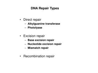
DNA Damage Tolerance and Repair 1-Dealing with Problems occurring during DNA replication • Mutations resulting from errors made during DNA replication Mismatch Repair Pathway • Ribonucleotides incorporated during DNA replication 2- Dealing with Problems coming after DNA replication resulting from DNA damage • Non exhaustive list of Damages • Strategies and mechanisms of DNA damage tolerance and repair: Bypass/Translesion DNA polymerases Direct Reversal Base Excision Repair Nucleotide Excision Repair • • Not treated: • Double-stranded break repair • Transcription coupled DNA repair
10-2 Editing 10-6 ? 10-8-10-9 DNA methylation 1) DNA methylation is delayed after replication 2) Dam methylase ! GATC New strand CTAG 3HC GATC CH3 CH3 CTAG CH3 +3HN COO- : NH2 N7 S+ A O N1 CH3 N9 N3 dR OH OH S-adenosyl methionine Increasing Replication Fidelity by mismatch repair ! ? New strand template How to differentiate between right and wrong Parental and new strand ?
Methyl-directed mismatch repair in prokaryotes MutL/MutS MutH CH3 Uvr genes = genes that promote UV Resistance DNA sliding through MutL/MutS ATP Mut genes = When these genes are mutated, bacteria show increased rates of mutations ADP+Pi CH3 ATP Ligation ADP+Pi CH3 Cleavage by MutH Filling the gap CH3 CH3 DNA unwinding by Helicase II (MutU/UvrD) CH3 CH3 Exonuclease VII (5’-->3’) or Exonuclease I (3’-->5’)
The Problem of ribonucleotide incorporation by DNA polymerases • DNA polymerases discriminate deoxyvsriboNTP, but not at 100% • [rNTPs] >> [ dNTPs] in vivo -> this leads to riboNTP incorporation in newly synthesized DNA a a e a a e e d e d d d (PNAS 107, p4950, 2010) Enzymes that deal with removal of RNA in Okazaki fragments also remove riboNTPsmis-incorporated into DNA : Rnase H, Fen1, DNA Pol.I
Post-replicative DNA Damages that need to be repaired or dealt with - Oxidative damages •G -> 8 oxoguanine •Strand Break • Pyrimidinedimers • (UV light) - Hydrolysis of glycosidic bond (Depurination) - Alkylation of bases Methylation of guanine N6 G-> O6meG - Deamination of bases •Spontaneous •Chemically induced •C->U •5 meC -> T •A->HX - Bulky DNA adducts
Induction of PyrimidineDimers by UV light PDB ID = 1SM5
Spontaneous Deaminations C --> U 10-7/24 hours: 100 events/day for a mammalian cell A --> H 10-9/24 hours G --> X 10-9/24 hours
The problem of Uracil in DNA A C A T G G T G U A U C 5’ 3’ 5’ 3’ Why Uracil was not selected as a natural base in DNA: Due to incorporation of dUTP by polymerase If Uracil were a natural base, the DNA repair machinery would not know whether these Uracil are “normal” uracil that come from incorporation by polymerase, or “non-natural” uracil coming from deamination of cytosines. Since there is no way to discri- minate between these, the genetic systems did not select uracil as a natural base (exceptions) Due to spontaneous Deamination of C->U
Spontaneous Depurinations 1/105 in 24 hours: 10,000 events/day for a mammalian cell
Oxidative damage of DNA O H N NH O N N NH2 8-oxo Guanine - Source of oxidative agents Consequences for Nucleotides: • Respiratory Chain: O CytC oxidase 2H2O O2 N H2O2 NH 1e- 2H+ -OH + .OH H2O2 O2- N -OH NH2 N 1e- Guanine • Fenton Chemistry (Metals) 5-formyl Uracil - Neutralization of reactive species Thymine superoxide 2O2-+2H+ H2O2 +O2 Deoxyribose Ribose dismutase catalase 2H2O2 2H2O+O2 No cellular Neutralization -> Main source of Oxidative agent .OH Consequences for Nucleic Acids: Strand Breaks (bad for DNA replication)
8-oxo-guanine generates replication block or G-C -> T:A transversions after DNA Replication DNA Polymerases tend to incorporate A opposite to 8-oxoG because of the tendency of 8-oxoG to switch in the syn conformation; A is the only nt that can form a base pair with syn8-oxoG whose geometry resembles that of a Watson-Crick base pair G-C H2O2 -OH 8xoG•C DNA replication 8xoG•A J Am Chem Soc. 2005 Oct 12;127(40):13906-18 8xoG•A T-A
Bulky DNA adducts caused by: diesel engine exhaust cigarette smoke block DNA Replication cooking/broiling of food
1- Bypass of lesions: avoids DNA replication stalls • bypass of DNA damage bytranslesionDNA Polymerases -> not a “repair” but is used to prevent DNA replication blocks 2- Direct Reversal of Damage (alkylation of bases, pyrimidinedimers) • Photolyase reversion of Y dimers • Dealkylation of guanines by suicidal MGMTase • Dealkylation of 1mA and 3mC by AlkB (not shown) DNA repair and tolerance Strategies & Enzymes 3- Base excision repair (deamination, alkylation, oxidation of bases) • Uracil-N glycosylase • 8-oxoG glycosylase 4- Nucleotide excision repair (pyrimidinedimers, bulky DNA adducts) • Bacteria: UvrA, UvrB, UvrC, Helicase II (UvrD) • DNA pol. I, DNA ligase • Eukaryotes : Xerodermapigmentosum proteins, TFIIH
Bypass of 8-oxoG lesions by a specialized eukaryotic DNA polymerase (Pol Pol d Pol X* X X X* <- Bypass product 75nt- X = Guanosine X * = 8-Oxo Guanosine Bacterial and Eukaryotic cells possess multiple translesion polymerases that are capable of bypassing DNA lesions Primer Extension Assay to map template replication by the two DNA polymerases -X* Block of polymerization at 8-oxoG Primer- Primer (41nt) 5’-AGG Template (75nt) 3’-TCCGTAXAATG--5’
Switch between Replicative and Translesion DNA polymerases involves PCNA Ubiquitination and prevents stalling of replication at the sites of DNA damage A DNA lesion (red) causes stalling of the replicative DNA Pol.d. PCNA 2) The E3 ubiquitinligase Rad18 guides Pol.h (aTLS DNA polymerase) to stalled replication forks. 3) Rad18 monoubiquitinates PCNA at stalled replication forks. PCNA (4) Pol.h binds to mono-ubiquitinated PCNA and performs replicative bypass of damaged DNA, preserving replication fork movement. PCNA http://www.pathology.unc.edu/faculty_labs/vaziri_lab/image012.jpg
Direct Reversal: h damage Repair of pyrimidinedimers DNA photolyase Photoreactivating enzyme with 2 chromophores h 300-500nm e.g. 5-deazaflavin (or N5N10methyleneTHF) FADH- Photoreactivation
Dealkylation of guanines by Methyl Guanine Methyl Transferase (MGMT) • Mutations of Human Homologues of O6MGMT linked to cancer : maintaining DNA information is required for tumor suppression • The “inactivated” enzyme serves as a transcription factor to induce expression of DNA repair genes -> amplifies the cellular response to DNA damage
damaged base Base Excision Repair:General strategy DNA glycosylase/ glycosidase Cleaves glycosidic bond at damaged base apurinicor apyrimidinicsite + AP endonuclease Cuts strand at AP site (APurinic or Apyrimidic) leaves 5’ terminal deoxyribose P moiety OH P Second cleavage (1nt further) by another enzyme OH P DNA Pol + DNA ligase Probably removes several nts by nick translation
Two types of base excision repair mechanisms Typically one enzyme cleaves the glycosidic bond of the damaged base, then the phosphodiester backbone is cleaved by an endonuclease specific for sites lacking a base (AP sites) Two sequential enzymatic activities are involved on two different enzymes: Pure glycosylase/glycosidase then B) AP endonuclease Ex: Uracil N-glycosylase cleaves glycosidic bonds of deoxyuridine but does not have AP endonuclease activity – needs another enzyme 2) The same polypeptide carries both glycosylase & AP endonuclease activities Ex: 8-OxoG DNA glycosylase (hOGG1)
Structure and activity of uracil N-glycosylase PDB ID = 2OXM
DNA Repair Strategies for 8-oxoG damage OGG1 = glycosidase for 8-oxoG MUTYH = glycosidase for A’s misincorporated in front of the 8-oxoG’s David et al., Nature447, 941 - 950 (2007)-Figure 1
How does hOGG1 recognize 8-OxoG damages ? recognition of 8-OxoG recognition of unpaired C The oxoG base is stacked between Phe 319 and Cys 253. Residues Gly 42, Gln 315 and two water molecules hydrogen bond to the Watson–Crick and Hoogsteen faces of the lesion base. The cytosine paired opposite oxoG is recognized by H-bonding Interactions with Arg 154 and Arg 204, and an additional H bond with Asn 149. PDB ID = 1EBM Bruner et al., Nature403, 859 - 866 (24 February 2000)-Figure 6
Nucleotide Excision Repair in Prokaryotes Uvr = UV Resistance When mutated, genes coding for Uvr proteins show an decrease in UV resistance
Nucleotide Excision Repair in Eukaryotes (Y dimer, bulky adducts) TFIIH = basal RNA Polymerase II transcription factor H; contains XPB & XPD subunits important because it links DNA repair to transcription XP = Xeroderma pigmentosum