Download
1 / 50
1.18k likes | 3.55k Views
DNA repair mechanisms. Dr.S.Chakravarty MBBS , MD. DNA Damage . During replication – corrected by proof reading Environmental factors : physical and chemical factors – corrected by DNA repair mechanisms. Already proof reading !! Why Repair ?. Learning objectives.

E N D
DNA repair mechanisms Dr.S.Chakravarty MBBS , MD
DNA Damage • During replication – corrected by proof reading • Environmental factors : physical and chemical factors – corrected by DNA repair mechanisms.
Learning objectives • Outline the Causes and Types of DNA damage that can occur in the living cell • Describe in detail the 4 types of DNA repair mechanisms • List the Key enzymes associated with DNA damage repair mechanisms and their defects
Types of DNA damage 1. Single base alteration : • Deamination of cytosine to uracil • Depurination • Demination of adenine to hypoxanthine Deamination of Cytosine to Uracil Mutations , wrong reading of the Genetic code
Two base alteration : • UV light induced thymine – thymidine dimers Adjacent Thymidine dimers
3. Chain breaks: • Ionizing radiation • Radioactive disintegration • Oxidative free radicals 4. Cross linkage : Chemicals - vinyl chloride and hydrogen peroxide, and Polycyclic aromatic hydrocarbons found in smoke
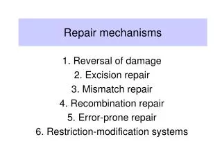
DNA repair • Mismatch repair • Base excision repair • Nucleotide excision repair • Double stranded break repair
Mismatch repair • Mismatch repair is strand specific. • G2 PHASE OF CELL CYCLE …CORRECTS DNA REPLICATION ERRORS • Mismatch repair machinery can distinguish between template and newly synthesized strands. HOW DOES THIS HAPPEN ? • An enzyme called as the DAM methylasemethylates DNA at N6 position for all adenines within 5’ GATC sequences • Newly synthesized strands are not methylated immediately. This allows to distinguish b/w the two.
Faulty mismatch repair • HNPCC – hereditary non polyposis colon cancer (LYNCH syndrome) • Autosomal Dominant • 60% due to defect in hMSH-2 gene or hMLH1 • Mutations happen in microsatellite repeat region leading to instability • Risk of other cancers (Liver) Microsatellites are di and tri nucleotide repeats throughout the genome in non coding sections.(Normal phenomenon) Microsatellite instability :- Cells which lack mismatch repair The number of microsatellites differ in the mutated cells
Base excision repair • Correction for Spontaneous deamination or depurination. • G1 phase of cell cycle • Identified by DNA glycosylase and removed. • The attached sugar and phosphate are removed by Apurinic or Apyrimidinic endonucleases. Harper’s Biochemistry 26th edition
Action of DNA glycosylases. These enzymes hydrolyze the glycosidic bond of their corresponding altered base (red)
Nucleotide excision repair • Mechanism used to replace DNA upto 30 base pairs. • Excision endonuclease (exinuclease) identifies and cleaves a fragment of DNA of around 20 -25 base pairs. • G1 phase of cell cycle
Nucleotide excision repair EXCISION ENDONUCLEASE (EXCINUCLEASE)
Xerodermapigmentosa • Autosomal recessive • Defect in nucleotide excision repair (XPA and XPC) gene • Extreme UV light sensitivity • Excessive freckling • Corneal ulcerations • Multiple skin cancers
Double strand break repair • Free radical and ionizing radiation cause double strand breaks. • Ku protein and DNA dependent protein kinaseare involved in repair. • It is also physiologically seen in immunoglobulin gene rearrangement.
Fanconi anemia • FA is the result of a genetic defect in a cluster of proteins responsible for DNA repair. • 1 per 350,000 births with a higher frequency in Ashkenazi Jews and Afrikaners in South Africa • Stunted growth • Skeletal deformities – digits • Café-au-lait spots • Myelodysplastic syndromes • Acute myelogenous leukemia • Bone marrow failure
Bloom syndrome • Characterized by a high frequency of breaks and rearrangements in an affected chromosomes. • Mutations in the BLMgene • Short stature, characteristic long face, butterfly shaped rash, micrognathia, hypogonadism
Ataxia telengiectasia • Defect in the ATM gene, responsible for recognizing and correcting errors in duplicating DNA when cells divide. • Cerebellar ataxia • Chromosomal instability • Telengiectasias of blood vessels • Immunodeficiency
Mechanism of DNA repair Type of Defect Repair mechsnism: Mismatch repair Base excision repair Nucleotide excision repair Double stranded break repair Copying errors during replication Spontaneous chemical or radiation damage to a single base Chemical or radiation to a segment Ionising radiation, chemotheray and free radicals
Autosomal recessive:F - Fanconi anemiaA - Ataxia telangiectasiaB - Bloom syndromeX - Xerodermapigmentosum Autosomal dominant:C- hereditary non-polyposisColorectal Cancer
Question 1 Patients with hereditary nonpolyposis colon cancer [HNPCC(114500)] have genes with microsatellite instability, that is, many regions containing abnormal, small loops of unpaired DNA. This is a result of a mutation affecting a. Mismatch repair b. Chain break repair c. Base excision repair d. Depurination repair e. Nucleotide excision repair
Question 2 Following ultraviolet damage of DNA in skin a. A specific excinuclease detects damaged areas b. Purinedimers are formed c. Both strands are cleaved d. Endonuclease removes the strand e. DNA hydrolysis does not occur
Question 3 Xerodermapigmentosum is an inherited human skin disease that causes a variety of phenotypic changes in skin cells exposed to sunlight. The molecular basis of the disease appears to be Rapid water loss caused by defects in the cell membrane permeability b. The inactivation of temperature-sensitive transport enzymes in sunlight c. The induction of a virulent provirus on ultraviolet exposure d. The inability of the cells to synthesize carotenoid-type compounds e. A defect in an excision-repair system that removes thymine dimers from DNA
Question 4 • Dyskeratosis congenital is a genetic condition with impaired proliferation of stem cells. The defect has been traced to an inadequate production of an enzyme required for chromosome duplication in nuclei of dividing cells. This enzyme contains a single stranded RNA.What is the deficient enzyme? • A) DNA POLYMEASE δ • B)DNA LIGASE • C)PRIMASE • D)TELOMERASE • E)DNA POLYMERASE γ
Learning Objectives • Briefly describe the stages of cell cycle and its check points • Explain the importance of P53 and Rb proteins in cell cycle regulation
Cell cycle CELLULAR DIVISION Interphase - Cell spends almost 90% of its time in this phase
How long does the cell cycle last? • Well, it depends on the cell type !! • Stem cells, embryonic cells- a few hours • Some cells divide very slowlyNeurons • Some cells divide when inducedLymphocytes
Stages of cell cycle • G0 stage : • Post mitotic stage • Quiescent or senescent stage • Permanent stage for fully differentiated cells: Ex: Neurons, Beta cells. • Interphase : G1 – Phase m RNA transcription S – Phase G2 – Phase Replicated DNA is assessed Protein synthesis Preparatory phase: Accumulation of Nutrients Replication phase
G1 phase • Ending of previous mitosis to beginning of DNA synthesis. • Increased biosynthetic activity of the cells • Rapid growth • Enzymes required for the S phase are synthesized. • Average Duration - 6-12 hours
S phase • DNA synthesis • Doubling of DNA content – replication • Low protein synthesis. • but histone production is normal. • Average Time – 6-8 hours
G2 phase • Microtubule formation: • Further protein and organelle synthesis • Average Time – 3-4 hrs M phase • Divided into Prophase, Metaphase, Anaphase, Telophase – karyokinesis • Cytokinesis – division of cytoplasm
Regulation of cell cycle • Cyclins - Regulatory subunits • Cyclin dependent kinases – catalytic subunits • phosphorylation of proteins which drives the cell cycle.
Cell cycle Control Cyclin D binds to existing CDK4, forming the active cyclin D-CDK4 complex. Cyclin D-CDK4 complex in turn phosphorylates the retinoblastoma susceptibility protein (Rb) hyperphosphorylatedRbdissociates from the E2F/DP1/Rbcomplex (which was bound to the E2F responsive genes, effectively "blocking" them from transcription), activating E2F G2/M check point M phase Rb Cyclin B CDK-1 (+) Cyclin D CDK 6,4 (+) G1 G2 Rb DISSOCIATES P (+) Cyclin E CDK-2 S (+) G1/S check point Cyclin A CDK-2
Inhibitors of cell cycle: Central role of p53 INK4a/ARF locus ARF tumor suppressor (-) (-) MDM-2 RbPhosphorylation (-) ATM gene (+) (+) p53 BAX, BAD, BIM (+) (+) (+) p21 p27 p57 Apoptosis Inhibition of cell cycle MDM -murine double minute ATM-Ataxia telangiectasia mutated
TUMOR SUPPRESSOR GENE P53 p53 : most common gene involved in cancers • Tumor suppressor gene altered in many cancers • Causes cell arrest and apoptosis • Stimulates apoptosis by stimulating BAX gene. • MDM-2 regulates p53 by feedback. • Main control point at G1/S checkpoint and component of G2/M checkpoint
Inhibitors of cell cycle CIP/KIP family (cyclin-dependent kinases inhibitors) INK4/ARF family Examples – p21,p27 and p57 Blocks the cell cycle by binding to Cyclin /CDK complexes. Activated by p53 – tumor suppressor gene Examples - p16 and p14 Promotes the inhibitory effects of Rb gene. Increase p53 levels by inhibiting MDM-2 protein.
ATM gene • Ataxia telengiectasia mutated gene: • Activated by DNA breaks in cell cycle • Acts through p53 at G1/S phase • At G2/M phase – Disrupts cyclin B –CDK-1 complex. • ATM additionally phosphorylates MDM2 and p53
Colorectal Tumorigenesis 1When Ras(Rat sarcoma) is 'switched on' by incoming signals, it subsequently switches on other proteins, which ultimately turn on genes involved in cell growth, differentiation and survivalunintended and overactive signalling inside the cell, even in the absence of incoming signals 2