Download
1 / 30
300 likes | 419 Views
Sperm Structure and Function. The head contains the nucleus and the enzyme-filled acrosome . Acrosome enzymes allow the sperm to penetrate the egg's barriers. The neck encloses a centriole. The centriole will fuse with a second centriole, contributed by the egg, to form the centrosome.

E N D
Sperm Structure and Function • The head contains the nucleus and the enzyme-filled acrosome. • Acrosome enzymes allow the sperm to penetrate the egg's barriers. • The neck encloses a centriole. • The centriole will fuse with a second centriole, contributed by the egg, to form the centrosome. • The midpiece is packed with mitochondria, which produce the ATP necessary for movement. • The tail has a flagellum that acts as a propeller.
Egg Structure and Function • Egg cells are relatively large (nutrient storage) and nonmotile. • Quantity of nutrients varies across species. • The relatively small mammalian egg only has to supply nutrients for early development, as embryos start to obtain nutrition through the placenta shortly following fertilization. • Egg-laying species produce larger eggs; the yolk of the egg is the embryo’s sole source of nutrition prior to hatching.
Other Egg Structures • The eggs of many species contain cytoplasmicdeterminants that control the early events of development. • Many eggs also contain corticalgranules, small enzyme-filled vesicles that are activated during fertilization. • The vitelline envelope,a fibrous, mat-like sheet of glycoproteins, surrounds the egg. • Mammals have an unusually thick vitelline envelope called the zonapellucida. • Some species have a jelly layer, a thick, gelatinous matrix around the vitelline envelope for further protection.
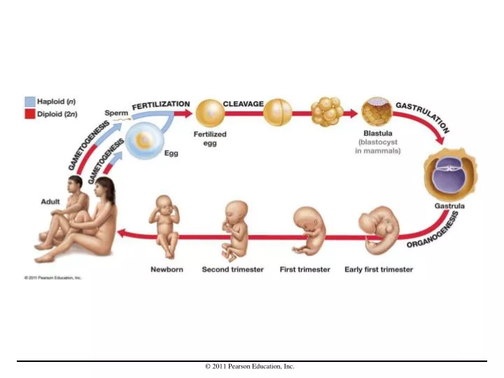
Fertilization • Fertilization occurs when a haploid sperm and egg cells fuse, forming a diploid zygote (a fertilized egg). • Many conditions must be met before a zygote can form: • Gametes must be in the same place at the same time. • Gametes must recognize and bind to each other. • Gametes must fuse together. • Fusion must trigger the onset of development. • Sea urchins are a model system for studying fertilization; they produce large numbers of gametes and undergo external fertilization.
Fertilization in Sea Urchins • When a sperm head contacts the jelly layer, enzymes from the sperm’s acrosome digest through the egg's jelly layer and vitelline envelope. • The plasma membranes of sperm and egg fuse when the sperm head contacts the surface of the egg cell. • Fertilization is complete when the sperm nucleus, mitochondria, and centriole enter the egg, and the sperm and egg nuclei fuse to form the zygote nucleus.
Gametes from the Same Species Recognize Each Other • Bindin is a protein on the head of sea urchin sperm that binds to the surface of sea urchin eggs. • Bindin acts as a “key,” such that a sperm binds only to eggs of the same species.
Identifying the Egg Cell Receptor for Sperm • By treating sea urchin egg cells with a protease (an enzyme that cleaves peptide bonds), researchers isolated the fragment of the egg cell receptor that binds to sperm. • Fertilizin is a compound on the surface of sea urchin egg cells that is involved in binding sperm. It acts as the “lock” to bindin’s “key.” • Each species has its own version of fertilizin and bindin, making the lock-and-key interaction both necessary for fertilization and species specific.
Why Does Only One Sperm Enter the Egg? • Animals employ different mechanisms to avoid polyspermy, fertilization by more than one sperm. • In sea urchins, fertilization stimulates the creation of a physical barrier. • After fertilization, a Ca2+-based signal is rapidly induced and propagated throughout the egg, resulting in the formation of a fertilization envelope, which keeps away additional sperm. • In mammals, the cortical granules release enzymes that modify egg cell receptors, preventing binding by additional sperm.
Cleavage • Cleavage • is the set of rapid cell divisions that take place in animal zygotes immediately after fertilization. • first step in embryogenesis, the process that makes a single-celled zygote into a multicellular embryo. • partitions the egg cytoplasm • The cells created by cleavage divisions are called blastomeres. • When cleavage is complete the embryo consists of a mass of blastomere cells called a blastula (in mammals: blastocyst).
What Role Do Cytoplasmic Determinants Play? • Cytoplasmic determinants are found in specific locations within the egg cytoplasm, so they end up in specific populations of blastomeres. • will result in differentiation of cells
Cleavage in Mammals • Cleavage occurs in the mammalian oviduct (fallopian tubes), which connects the ovary, where the egg matures, to the uterus, where the embryo develops. • Cleavage results in a blastocyst, a specialized blastula consisting of two populations of cells: • The external, thin-walled hollow trophoblast surrounds the inner cell mass (ICM). • After the blastocyst embeds in the uterine wall, a mixture of trophoblast and maternal cells form the placenta, an organ which provides nourishment and waste removal for the developing embryo. • The ICM contains the cells that undergo gastrulation and develop into the embryo.
Gastrulation • During gastrulation, extensive and highly organized cell movements radically rearrange the embryonic cells into a structure called the gastrula. • Gastrulation results in the formation of embryonic tissue layers. A tissue is an integrated set of cells that function as a unit. • Most early embryos have three primary tissue layers: ectoderm, mesoderm, and endoderm. • These embryonic tissues are called germlayers because they give rise to adult tissues and organs.
Gastrulation in Frog Embryos • The frog blastula contains a fluid-filled space called the blastocoel. • Gastrulation begins with the formation of an opening called a blastopore. • Cells from the periphery move inward through the blastopore, forming a tube-like structure that will become the gut.
Germ Layers • Ectoderm forms the outer covering of the adult body and the nervous system. • Mesoderm gives rise to muscle, most internal organs, and connective tissues such as bone and cartilage. • Endoderm produces the lining of the digestive tract or gut, along with some of the associated organs.
The Completion of Gastrulation • In addition to establishing the embryonic tissue layers, gastrulation has another major outcome: The major body axes become visible. • At the end of gastrulation, the three embryonic tissues are arranged in layers, the gut has formed, and the major body axes have become visible.
Organogenesis • Organogenesis is the process of tissue and organ formation that begins once gastrulation is complete and the embryonic germ layers are in place. • During organogenesis, cells proliferate and become differentiated, meaning that they become a specialized cell type. • Differentiated cells have a distinctive structure and function because they express a distinctive suite of genes.
Organizing the Mesoderm into Somites • Early in organogenesis, the rod-like notochord appears in the dorsal mesoderm. • notochord characterizes chordates, which includes humans and other vertebrates. • The notochord functions as a key organizing element during organogenesis. • In many chordates, as organogenesis continues, the notochord cells undergo apoptosis.
Neural Tube Formation • Signals from the notochord trigger reorganization of the dorsal ectodermal cells, leading to neural tube formation. • The neural tube is the precursor to the brain and spinal cord.
Somite Formation • Once the neural tube forms, the mesodermal cells become organized into blocks of tissues called somites, which form on both sides of the neural tube down the length of the body.
Somite Formation • Once the neural tube forms, the mesodermal cells become organized into blocks of tissues called somites, which form on both sides of the neural tube down the length of the body.
Somite Maturation and Determination • Somite cells form a variety of structures, but are initially not determined, meaning they can become any of the somite-derived elements of the body. • As the somite matures, somite cells becomeirreversibly determined, and will eventually differentiate into a specific cell type based on their location within the somite.
The Process of Determination • In the process of determination, somite cells differentiate in response to signals from nearby tissues. • These signals diffuse away from cells in the notochord, the neural tube, and nearby ectoderm and mesoderm to act on specific populations of target cells in the somite.