Download
1 / 1
10 likes | 264 Views
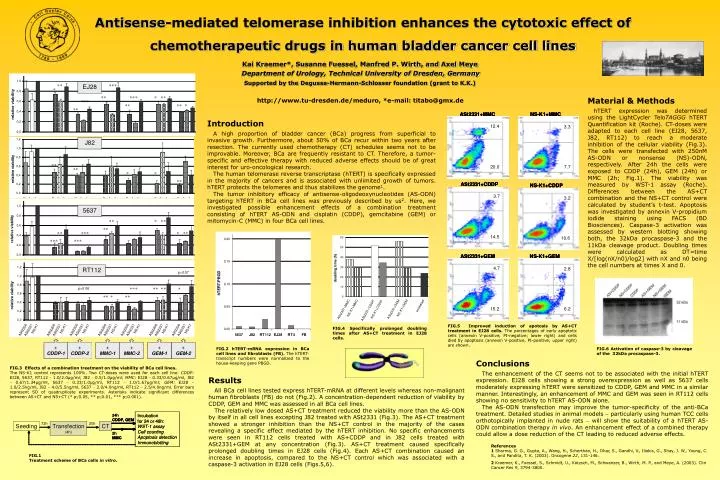
Antisense-mediated telomerase inhibition enhances the cytotoxic effect of chemotherapeutic drugs in human bladder cancer cell lines. Kai Kraemer*, Susanne Fuessel, Manfred P. Wirth, and Axel Meye Department of Urology, Technical University of Dresden, Germany

E N D
Antisense-mediated telomerase inhibition enhances the cytotoxic effect ofchemotherapeutic drugs in human bladder cancer cell lines Kai Kraemer*, Susanne Fuessel, Manfred P. Wirth, and Axel Meye Department of Urology, Technical University of Dresden, Germany Supported by the Degussa-Hermann-Schlosser foundation (grant to K.K.) Material & Methods hTERT expression was determined using the LightCycler TeloTAGGG hTERT Quantification kit (Roche). CT-doses were adapted to each cell line (EJ28, 5637, J82, RT112) to reach a moderate inhibition of the cellular viability (Fig.3). The cells were transfected with 250nM AS-ODN or nonsense (NS)-ODN, respectively. After 24h the cells were exposed to CDDP (24h), GEM (24h) or MMC (2h; Fig.1). The viability was measured by WST-1 assay (Roche). Differences between the AS+CT combination and the NS+CT control were calculated by student’s t-test. Apoptosis was investigated by annexin V-propidium iodide staining using FACS (BD Biosciences). Caspase-3 activation was assessed by western blotting showing both, the 32kDa procaspase-3 and the 11kDa cleavage product. Doubling times were calculated as DT=time X/[log(nX/n0)/log2] with nX and n0 being the cell numbers at times X and 0. http://www.tu-dresden.de/meduro, *e-mail: titabo@gmx.de Introduction A high proportion of bladder cancer (BCa) progress from superficial to invasive growth. Furthermore, about 50% of BCa recur within two years after resection. The currently used chemotherapy (CT) schedules seems not to be improvable. Moreover, BCa are frequently resistant to CT. Therefore, a tumor-specific and effective therapy with reduced adverse effects should be of great interest for uro-oncological research. The human telomerase reverse transcriptase (hTERT) is specifically expressed in the majority of cancers and is associated with unlimited growth of tumors. hTERT protects the telomeres and thus stabilizes the genome1. The tumor inhibitory efficacy of antisense-oligodeoxynucleotides (AS-ODN) targeting hTERT in BCa cell lines was previously described by us2. Here, we investigated possible enhancement effects of a combination treatment consisting of hTERT AS-ODN and cisplatin (CDDP), gemcitabine (GEM) or mitomycin-C (MMC) in four BCa cell lines. FIG.5 Improved induction of apotosis by AS+CT treatment in EJ28 cells. The percentages of early apoptotic cells (annexin V-positive, PI-negative; lower right) and cells died by apoptosis (annexin V-positive, PI-positive; upper right) are shown. FIG.4 Specifically prolonged doubling times after AS+CT treatment in EJ28 cells. FIG.2 hTERT-mRNA expression in BCa cell lines and fibroblasts (FB).The hTERT-transcript numbers were normalized to the house-keeping gene PBGD. FIG.6 Activation of caspase-3 by cleavage of the 32kDa procaspase-3. Conclusions The enhancement of the CT seems not to be associated with the initial hTERT expression. EJ28 cells showing a strong overexpression as well as 5637 cells moderately expressing hTERT were sensitized to CDDP, GEM and MMC in a similar manner. Interestingly, an enhancement of MMC and GEM was seen in RT112 cells showing no sensitivity to hTERT AS-ODN alone. The AS-ODN transfection may improve the tumor-specificity of the anti-BCa treatment. Detailed studies in animal models particularly using human TCC cells orthotopically implanted in nude rats will show the suitability of a hTERT AS-ODN combination therapy in vivo.An enhancement effect of a combined therapy could allow a dose reduction of the CT leading to reduced adverse effects. FIG.3 Effects of a combination treatment on the viability of BCa cell lines. The NS-K1 control represents 100%. Two CT-doses were used for each cell line. CDDP: EJ28, 5637, RT112 1.0/2.0µg/ml; J82 0.5/1.0µg/ml; MMC: EJ28 0.33/0.67µg/ml, J82 0.67/1.34µg/ml, 5637 0.33/1.0µg/ml, RT112 1.0/1.67µg/ml; GEM: EJ28 1.0/2.5ng/ml, J82 4.0/5.5ng/ml, 5637 2.0/4.0ng/ml, RT112 2.5/4.0ng/ml. Error bars represent SD of quadruplicate experiments. Asterisks indicate significant differences between AS+CT and NS+CT (* p0.05, ** p0.01, *** p0.001). Results All BCa cell lines tested express hTERT-mRNA at different levels whereas non-malignant human fibroblasts (FB) do not (Fig.2). A concentration-dependent reduction of viability by CDDP, GEM and MMC was assessed in all BCa cell lines. The relatively low dosed AS+CT treatment reduced the viability more than the AS-ODN by itself in all cell lines excepting J82 treated with ASt2331 (Fig.3). The AS+CT treatment showed a stronger inhibition than the NS+CT control in the majority of the cases revealing a specific effect mediated by the hTERT inhibition. No specific enhancements were seen in RT112 cells treated with AS+CDDP and in J82 cells treated with ASt2331+GEM at any concentration (Fig.3). AS+CT treatment caused specifically prolonged doubling times in EJ28 cells (Fig.4). Each AS+CT combinationcaused an increase in apoptosis, compared to the NS+CT control which was associated with a caspase-3 activation in EJ28 cells (Figs.5,6). References 1 Sharma, G. G., Gupta, A., Wang, H., Scherthan, H., Dhar, S., Gandhi, V., Iliakis, G., Shay, J. W., Young, C. S., and Pandita, T. K. (2003). Oncogene 22, 131-146. 2Kraemer, K., Fuessel, S., Schmidt, U., Kotzsch, M., Schwenzer, B., Wirth, M. P., and Meye, A. (2003). Clin Cancer Res 9, 3794-3800. FIG.1 Treatment scheme of BCa cells in vitro.