Download
1 / 20
240 likes | 552 Views
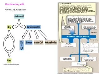
BIOCHEMISTRY BONE METABOLISM MSK BLOCK SYSTEM. Nabil Bashir October 1 st , 2009. Bone. Inorganic (67%) Hydroxyapatite 3 Ca 10 (PO 4 ) 6 (OH) 2 There is some amorphous calcium phosphate Organic (33%) component is called osteoid Type I collagen (28%) Non-collagen structural proteins (5%)

E N D
BIOCHEMISTRYBONE METABOLISMMSK BLOCK SYSTEM Nabil Bashir October 1st, 2009
Bone • Inorganic (67%) • Hydroxyapatite 3 Ca10(PO4)6(OH)2 • There is some amorphous calcium phosphate • Organic (33%) component is called osteoid • Type I collagen (28%) • Non-collagen structural proteins (5%) • Proteoglycans • Sialoproteins • Gla-containing proteins (gamma carboxyglutamate) • Phosphoproteins • Bone specific proteins: osteocalcin, osteonectin • Growth factors and cytokines (Trace) • Bone undergoes continuous turnover or remodeling throughout life • About 20% of bone is undergoing remodeling at any one time
Osteoblasts Bone formation Synthesis of matrix proteins Type I collagen Osteocalcin Others Mineralization Activation of osteoclasts via RANKL production Osteoclasts Bone resorption Degradation of proteins by enzymes Acidification RANK is activated by RANKL, and this leads to cells differentiation to osteoclasts Osteoblast and Osteoclast Function
Osteoclastogenesis: RANK/RANKL/OPG axis RANK: Receptor activator of nuclear factor (NF)-kB RANKL: RANK ligand OPG: Osteoprotegerin (cytokine) Activating factors: M-CSF, IL-6etc. ASBMR Bone Curriculum
Osteoclastogenesis Activating factors cause the lining cells to produce RANKL, and then the RANK of the preosteoclasts binds with the RANKL and the forms multinucleated activated osteoclasts (10-20 fused cells, called polykaryons) OPG also binds with RANKL, preventing the preosteoclast RANK from binding ASBMR Bone Curriculum
Hormonal Control of Resorption: Pro-resorptive Most of the proresorptive factors upregulate mRNA expression of RANKL in osteoblasts Boyle et al, Nature 2003
Hormonal Control of Resorption: Anti-resorptive Boyle et al, Nature 2003
Genetic Mutations Boyle et al, Nature 2003 What would an OPG knock-out mouse look like?
CALCIUM& PHOSPHORUS HOMEOSTASIS • PTH, • VIT D, • CALCITONIN, • ESTROGENS
Ca2+ Resorption E2 PO4 PO4 CT Ca2+ Mineralization PTH Reabsorption Ca2+ PO4 Bioactivation 25D 1,25D PTH BONE BLOOD 1,25 D PTH 1,25 D Absorption CaPO4 PTH 1,25 D INTESTINE 1,25 D Figure 1. Control of blood Ca2+ and PO43- matrix KIDNEY calcitonin (CT) can counteract the effect of PTH on bone resorption estrogen (E2) counteracts effects of PTH and 1,25(OH)2D3 on bone resorption 1,25(OH)2D3 increases intestinal and renal absorption of phosphate to help promote bone mineralization PTH, in response to low serum Ca, increases plasma Ca by increasing bone resorption, and renal reabsorption of Ca 1,25(OH)2D3, in response to low serum Ca, increases plasma Ca by increasing intestinal absorption, bone resorption, and renal reabsorption of Ca PTH prevents hyperphosphatemia, which could be caused by the PTH effect on bone resorption, by inhibiting renal reabsorption of phosphate PTH activates the hydroxylation of 25(OH)D3 to the active 1,25(OH)2D3 form
Bone Remodeling • Osteoclasts dissolve bone • Large multinucleated giant cells • Osteoblasts produce bone • Have receptors for PTH, CT, Vitamin D, cytokines, and growth factors • Main product is collagen • When osteoblasts become encased in bone, they become osteocytes
IL-6 IL-6; other cytokines activation ODF PTH osteoclast c-FMS receptor M-CSF Figure 3. Control of bone remodeling by PTH Osteoblast Bone constructor Osteoclast Bone destructor cAMP PKA Gs PTH ODF – osteoclast differentiating factor osteoblast Osteoprotegerin receptor decoy ODF receptor MOP MOP – monocytic osteoclast progenitor cells Differentiation and fusion M-CSF – macrophage colony stimulating factor
Figure 3. Control of bone remodeling by PTH and calcitonin Osteoblast Bone constructor Osteoclast Bone destructor cAMPPKA Gs PTH IL-6 IL-6; other cytokines inactivation CT Gs PKA cAMP activation ODF Calcitonin secreted by thyroid C-cells in response to hypercalcemia CT gene can yield calcitonin gene-related peptide (CGRP)if processed differently (alternative mRNA splicing) CGRP = a potent vasodilator
Kidney 1-OHase Liver 25-OHase OH OH Diet HO HO HO OH Vitamin D3 25(OH) D3 1,25(OH)2 D3 (active hormone form) 24-OHase (kidney; many other tissues) UV from sunlight High Ca2+ or PO43- Skin OH OH 7 HO 24,25(OH)2 D3 (inactive form) Provitamin D3 (7-dehydrocholesterol: Intermediate in cholesterol synthesis) HO Specific receptors in intestine, bone, kidney Ca: Intestinal absorption Renal reabsorption Bone resorption PO4: Intestinal absorption Renal reabsorption Figure 4. Photobiosynthesis of vitamin D3 and its metabolism
IL-6 IL-6; other cytokines inactivation CT Gs PKA cAMP activation ODF Osteocalcin osteoblast 1,25D3 ODF receptor Gla Ca2+ Differentiation and fusion osteoclast c-FMS receptor M-CSF Osteoblast Bone constructor Osteoclast Bone destructor VDR receptor Nucleus RXR receptor mRNA 1,25D3 Chemotaxis MOP Figure 7. Control of bone remodeling by 1,25(OH)2D3
Estrogen (E2) and Androgen Estrogen (E2) and Androgen E2 proliferation collagen syn. Collagen matrix Ca2+/PO43- Bone mineralization Osteoblast Bone constructor Osteoclast Bone destructor cAMPPKA Gs PTH IL-6 IL-6; other cytokines inactivation mRNA CT Gs 3 PKA cAMP activation ODF Osteocalcin 1,25D3 osteoblast PTH, 1,25D3 ODF receptor Gla Ca2+ MOP Osteoblast Differentiation and fusion osteoclast c-FMS receptor Figure 8. Control of bone remodeling by PTH and 1,25(OH)2D3 and antagonism of their effects by estrogen and androgen M-CSF