Download
1 / 47
490 likes | 799 Views
MR Signal Generation. FMRI Undergraduate Course (PSY 181F) FMRI Graduate Course (NBIO 381, PSY 362) Dr. Scott Huettel, Course Director. Housekeeping Clarifications. Updated Syllabus (on BlackBoard) Minor changes to the orders of labs and project planning later in the month

E N D
MR Signal Generation FMRI Undergraduate Course (PSY 181F) FMRI Graduate Course (NBIO 381, PSY 362) Dr. Scott Huettel, Course Director FMRI – Week 2 – MR Signal Scott Huettel, Duke University
Housekeeping Clarifications • Updated Syllabus (on BlackBoard) • Minor changes to the orders of labs and project planning later in the month • Please complete self-assessment questions weekly • Email to me (with “SAQ”, “Ch1” in the title) • These are not graded except for completion FMRI – Week 2 – MR Signal Scott Huettel, Duke University
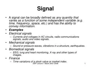
Outline for Today • Lecture: MR Signal Generation • Protons and the NMR property • Protons in a magnetic field: Alignment, Precession • Excitation and resonance • Reception and relaxation • Laboratory: Introducing MRI / fMRI data • Basic MATLAB use • Properties of MRI data • Basic neuroanatomy FMRI – Week 2 – MR Signal Scott Huettel, Duke University
Synopsis of MRI M: Put subject in strong magnetic field R: Transmit radio waves into subject, turn off transmitter, receive radio waves emitted by subject’s brain. This is the MR signal. I: Modulate the strength of the magnetic field slightly over space (next week). FMRI – Week 2 – MR Signal Scott Huettel, Duke University
1. Protons and the NMR property FMRI – Week 2 – MR Signal Scott Huettel, Duke University
Properties of Atomic Nuclei • Nuclei have two properties: • Spin (incompletely understood) • Charge (property of protons) • Nuclei are made of protons and neutrons • Both have spin values of ½ • Protons give charge • Pairs of spins tend to cancel, so only atoms with an odd number of protons or neutrons have spin A nucleus has the NMR Property if it has both angular momentum and a magnetic moment. Such nuclei have an odd number of protons or an odd number of neutrons. FMRI – Week 2 – MR Signal Scott Huettel, Duke University
The spinning mass of the proton generates an angular momentum J. The electric charge on the surface of the proton creates a small current loop, which generates magnetic momentμ. Both μ and J are vectors that point along the spin axis and whose direction is given by the right hand rule. FMRI – Week 2 – MR Signal Scott Huettel, Duke University
What nuclei can we measure? • Most common in our bodies: • 12C • 16O • 1H • 14N • Of these, only Hydrogen has the NMR property. • But, Hydrogen is the most abundant atom in the body • Mostly in water (H2O) This means that nearly all forms of MRI are measuring properties of Hydrogen. FMRI – Week 2 – MR Signal Scott Huettel, Duke University
2. Protons in a magnetic field FMRI – Week 2 – MR Signal Scott Huettel, Duke University
M FMRI – Week 2 – MR Signal Scott Huettel, Duke University
Protons in no magnetic field In the absence of a strong magnetic field, the spins are oriented randomly. Thus, there is no net magnetization (M). FMRI – Week 2 – MR Signal Scott Huettel, Duke University
Introduction of a Magnetic Field (B) Bo Image from G.A. Glatzmaier Computer simulation of Earth’s magnetic field (~0.3-0.6 Gauss, or 0.00006T) Helmholtz Pair Solenoid FMRI – Week 2 – MR Signal Scott Huettel, Duke University
Some Terminology Bo Bo Longitudinal Axis (z direction) B is used for magnetic fields. B0 is the scanner’s main field. Transverse Plane (xy plane) FMRI – Week 2 – MR Signal Scott Huettel, Duke University
Alignment with a magnetic field FMRI – Week 2 – MR Signal Scott Huettel, Duke University
Protons align with a magnetic field… FMRI – Week 2 – MR Signal Scott Huettel, Duke University
… but move around the field axis in a motion known as precession. Precession axis FMRI – Week 2 – MR Signal Scott Huettel, Duke University
In a magnetic field, protons can take high- or low-energy states FMRI – Week 2 – MR Signal Scott Huettel, Duke University
The difference between the numbers of protons in the high-energy and low-energy states results in a net magnetization (M). FMRI – Week 2 – MR Signal Scott Huettel, Duke University
Energy states: Temperature effects Low-energy protons at room temperature in Earth’s B: ~50.000000001% High-energy protons at room temperature in Earth’s B: ~49.999999999% Protons move back and forth between states because of thermal energy. As temperature decreases to near absolute zero, all protons move to lower-energy state. FMRI – Week 2 – MR Signal Scott Huettel, Duke University
When the magnetic field is weak, little energy is required for a proton to change between high and low states (ΔE is small). But, when the magnetic field is strong, much more energy is required (ΔE is large). Thus, protons in the lower-energy state tend to stay in that state Energy states: Magnetic field effects FMRI – Week 2 – MR Signal Scott Huettel, Duke University
The net magnetization (M) increases with increasing field strength (B0), but decreases with increasing temperature (T). FMRI – Week 2 – MR Signal Scott Huettel, Duke University
3. Excitation and Resonance FMRI – Week 2 – MR Signal Scott Huettel, Duke University
R FMRI – Week 2 – MR Signal Scott Huettel, Duke University
Excitation: Conceptual Overview FMRI – Week 2 – MR Signal Scott Huettel, Duke University
Key Concept: To measure magnetization we must perturb it • Protons must absorb energy to change between states • Parallel (aligned with) to B0 is lowest-energy state • Anti-parallel (aligned against) to B0 is highest-energy state • We can apply energy as electromagnetic radiation • Higher frequency radiation more energy • How can we calculate how much energy (i.e., at what frequency) to apply? FMRI – Week 2 – MR Signal Scott Huettel, Duke University
M (net magnetization) M (net magnetization) θ B1 (another very strong field) Let’s try adding another magnetic field… B0 (main field of scanner) FMRI – Week 2 – MR Signal Scott Huettel, Duke University
So, we could reorient some of the protons (i.e., change the net magnetization) by introducing a second, very strong magnetic field. Why is this impractical? FMRI – Week 2 – MR Signal Scott Huettel, Duke University
The angular momentum (J) is the product of the proton’s mass (m), it’s velocity (v), and its radius (r). Cool Fact #1: The current flow (I) and velocity (v) are vectors in the same direction (i.e., the charge is spinning just like the mass). Cool Fact #2: The rotation radius (r) and area (A) are proportional. The magnetic moment (μ) is given by the rotational force experienced by the proton (torque, or τmax) divided by the strength of the magnetic field. These are proportional to the moving charge of the proton (I) times the area around which its charge moves (A). Magnetic Moment and Angular Momentum have a constant relation! FMRI – Week 2 – MR Signal Scott Huettel, Duke University
If we assume that the proton is a point charge spinning in a circle, then the gyromagnetic ratio is given by a very simple equation (see book for derivation). Magnetic Moment and Angular Momentum have a constant relation! The constant γ (gamma) is known as the gyromagnetic ratio. It is fixed for any given atomic nucleus. The gyromagnetic ratio (γ) depends on only two things: charge (q) and mass (m). That’s it. FMRI – Week 2 – MR Signal Scott Huettel, Duke University
The gyromagnetic ratio (γ) is critical for MRI. It allows us to calculate the energy (expressed in electromagnetic frequency, v) needed to change an atomic nucleus from the low- to high-energy states in a given magnetic field (B0). This frequency (v) is known as the Larmor Frequency. It is the same as the precession frequency of the nucleus! FMRI – Week 2 – MR Signal Scott Huettel, Duke University
The Canonical Analogytm for resonance: a swing set • Option #1 • A single, strong push to lift the person off the ground • Requires an enormous amount of exertion, delivered very rapidly • Option #2 • Many small pushes at the resonant frequency of the swing set • Allows distribution of the energy over time! This is a random illustrative photo. The internet is great. FMRI – Week 2 – MR Signal Scott Huettel, Duke University
… moving from happy kids to atomic nuclei • Option #1 • Using a very strong perpendicular field • Impractical (perhaps impossible) to do quickly in a real device • Option #2 • Many small pushes at the resonant frequency of the atomic nucleus of interest • Allows distribution of the energy over time! Giving energy for a longer time period increases the flip angle. FMRI – Week 2 – MR Signal Scott Huettel, Duke University
“Tipping” in a rotating frame of reference The RF energy is called B1 because it is, in essence, a second magnetic field. FMRI – Week 2 – MR Signal Scott Huettel, Duke University
Resonance Frequencies of Common Nuclei Remember, the resonant frequency is constant for a given atomic nucleus and proportional to magnetic field strength. FMRI – Week 2 – MR Signal Scott Huettel, Duke University
What are the consequences of electromagnetic energy at this frequency? X-Ray, CT MRI (e.g., 6 x 10⁷Hz) MRI uses electromagnetic energy in the radio wave portion of the electromagnetic spectrum. It can cause heating of biological tissue, but does not break molecular bonds. FMRI – Week 2 – MR Signal Scott Huettel, Duke University
Radiofrequency Coils for Excitation (and Reception) • Defined by their function: Transmit / receive coil (most common) Transmit only coil (can only excite the system) Receive only coil (can only receive MR signal) • Defined by geometry Volume coil (low sensitivity but uniform coverage) Surface coil (High sensitivity but limited coverage) Phased-array coil (High sensitivity, near-uniform coverage) FMRI – Week 2 – MR Signal Scott Huettel, Duke University
4. Reception and relaxation FMRI – Week 2 – MR Signal Scott Huettel, Duke University
Tipping the net magnetization provides measurable MR signal! During Excitation (to) During Excitation (t1) The amount of current oscillates at the (Larmor) frequency of the net magnetization. Before Excitation After Excitation Excitation tips the net magnetization (M) down into the transverse plane, where it can generate current in detector coils (i.e., via induction). FMRI – Week 2 – MR Signal Scott Huettel, Duke University
Relaxation: Nothing Lasts Forever • Once we stop applying energy, M will go back to being aligned with static field B0 • This process is called relaxation • The part of M perpendicular to B0shrinks [Mxy] • This part of M is called transverse magnetization • It provides the detectable RF signal • Part of M parallel to B0 grows back [Mz] • This part of Mis called longitudinal magnetization • Mzis not directly detectable, but can be again converted into transverse magnetization by energy (e.g., B1) FMRI – Week 2 – MR Signal Scott Huettel, Duke University
T1 T2 FMRI – Week 2 – MR Signal Scott Huettel, Duke University
Relaxation Times and Rates • Net magnetization changes in an exponential fashion • Constant rate (R) for a given tissue type in a given magnetic field • R = 1/T, leading to equations like e–Rt • T1 (recovery): Relaxation of Mback to alignment with B0 • Usually 500-1000 ms in the brain (lengthens with bigger B0) • T2 (decay): Loss of transverse magnetization over a microscopic region ( 5-10 micron size) • Usually 50-100 ms in the brain (shortens with bigger B0) • T2*: Overall decay of the observable RF signal over a macroscopic region (millimeter size) • Usually about half of T2 in the brain (i.e., faster relaxation) FMRI – Week 2 – MR Signal Scott Huettel, Duke University
T1 and T2 parameters By selecting appropriate pulse sequence parameters (Week 4’s lecture), images can be made sensitive to tissue differences in T1, T2, or a combination. FMRI – Week 2 – MR Signal Scott Huettel, Duke University
T1 and T2 values at 1.5T FMRI – Week 2 – MR Signal Scott Huettel, Duke University
What about “I”? FMRI – Week 2 – MR Signal Scott Huettel, Duke University
I We just have signal, so far. We need spatial gradients to generate images. Next week. FMRI – Week 2 – MR Signal Scott Huettel, Duke University
MR Signal Generation • Scanners use very strong static fields (Tesla range) to generate net magnetization (M) • Protons in magnetic fields precess around the longitudinal axis of a field at the Larmor Frequency • Electromagnetic energy, when supplied at the Larmor frequency (radio waves) by head coils, is absorbed by the protons • This tips the net magnetization down into the transverse plane • As the net magnetization rotates through the transverse plane, it induces a changing current in the head coils. • This current is the MR signal FMRI – Week 2 – MR Signal Scott Huettel, Duke University
Laboratory #1 • Computers will be brought to our room • Form groups of 2 • Try to identify what other days/times work for your lab group • We need to coordinate on a couple of sessions • All lab exercises available on BlackBoard • You can print using EPrint FMRI – Week 2 – MR Signal Scott Huettel, Duke University