Download
1 / 29
420 likes | 1.1k Views
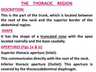
A-THE THORACIC WALL. A-THE THORACIC WALL. Boundaries. Boundaries. Posteriorly by the thoracic part of the vertebral column. Posteriorly by the thoracic part of the vertebral column. Anteriorly by the sternum and costal cartilages . Anteriorly

E N D
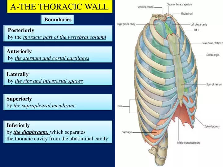
A-THE THORACIC WALL A-THE THORACIC WALL Boundaries Boundaries Posteriorly by the thoracic part of the vertebral column Posteriorly by the thoracic part of the vertebral column Anteriorly by the sternum and costal cartilages Anteriorly by the sternum and costal cartilages Laterally by the ribs and intercostal spaces Laterally by the ribs and intercostal spaces Superiorly by the suprapleural membrane Superiorly by the suprapleural membrane Inferiorly by the diaphragm, which separates the thoracic cavity from the abdominal cavity Inferiorly by the diaphragm, which separates the thoracic cavity from the abdominal cavity
1- STERNUM • It is a flat bone • Divides into three parts: 1-Manubrium sterni 2-Body of the sternum 3- Xiphoid process
The sternal angle (angle of Louis) formed by the articulation of the manubrium with the body of the sternum Lies at the level of second costal cartilage The point from which all costal cartilages and ribs are counted
2-Ribs There are 12 pairs of ribs, all of which are attached posteriorly to the thoracic vertebrae. The ribs are divided into three categories according to their relation to the sternum: True ribs: The upper seven pairs are attached anteriorly to the sternum by their costal cartilages False ribs: The 8th, 9th, and 10th pairs of ribs are attached anteriorly to each other and to the 7th rib by means of their costal cartilages. Floating ribs: The 11th and 12th pairs have no anterior attachment
Typical Rib A typical rib is a long, twisted, flat bone having a rounded, smooth superior border and a sharp, thin inferior border The inferior border forms THE COSTAL GROOVE which accommodates the intercostal vessels and nerve. intercostal vein intercostal artery intercostal nerve VAN important A rib has a head, neck, tubercle, shaft, and angle
3-The Vertebral Column is composed of 33 vertebrae 7 cervical 12 thoracic 5 lumbar 5 sacral (fused to form the sacrum) 4 coccygeal (the lower 3 are commonly fused)
A typical thoracic vertebra consists of: 1-a rounded body anteriorly (body bearing) 2-a vertebral arch posteriorly. (protect the spinal cord) They enclose a space called The vertebral foramen through which run the spinal cord and its coverings The vertebral arch gives rise to seven processes: a-One spinous b-Two transverse c- Four articular (2 superior 2 inferior)
Characteristics of a Typical Thoracic Vertebra • The body is heart shaped • The vertebral foramen is small and circular • The spines are long and inclined downward • Costal facets are present on the sides of the bodies for articulation with the heads of the ribs • Costal facets are present on the transverse processes for articulation with the tubercles of the ribs
The body and the vertebral arch are connected by means of pedicles
The pedicles • are notched on their • upper and lower borders • Forming • the superior and inferior • vertebral notches. On each side the superior notch of one vertebra and the inferior notch of an adjacent vertebra together form an intervertebral foramen. These foramina, in an articulated skeleton, serve to transmit the spinal nerves and blood vessels.
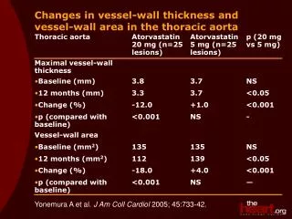
4-The diaphragm The diaphragm is a thin muscular and tendinous septum that separates the chest cavity above from the abdominal cavity below • The diaphragm is the most important muscle of respiration. • It is dome shaped and consists of a • peripheral muscular part • and a centrally placed tendon
Main Openings in the diaphragm The inferior vena cava passes through the central tendon at approximately vertebral level T8 The esophagus passes through the muscular part of the diaphragm, approximately at vertebral level T10 The aorta passes behind the posterior attachment of the diaphragm at vertebral level T12
Nerve supply of the diaphragm The phrenic nerves
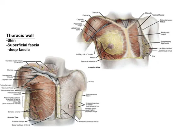
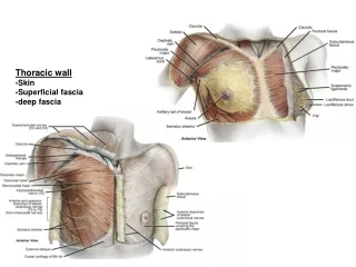
5-Intercostal Spaces 1-SKIN 2-SUPERFISCIAL FASCIA 3- THREE MUSCLES OF RESPIRATION: THE EXTERNAL INTERCOSTAL THE INTERNAL INTERCOSTAL THE INNERMOST INTERCOSTAL MUSCLE 4-THE ENDOTHORACIC FASCIA 5-THE PARIETAL PLEURA. The intercostal nerves and blood vessels run between the intermediate (internal intercostal) and deepest layers (innermost intercostal) of muscles They are arranged in the following order from above downward: INTERCOSTAL VEIN INTERCOSTAL ARTERY INTERCOSTAL NERVE (VAN)
The external intercostal muscle • the most superficial layer. • Its fibers are directed • downward and forward The Internal Intercostal Muscle • forms the intermediate layer. • Its fibers are directed • downward and backward
The innermost intercostal muscle Forms the deepest layer It is an incomplete muscle layer and crosses more than one intercostal space within the ribs.
B-CHEST CAVITY The chest cavity is bounded by the chest wall and below by the diaphragm
The chest cavity can be divided into MEDIAN PARTITION CALLED THE MEDIASTINUM LATERALLY PLACED PLEURAE AND LUNGS
2-Pleurae The pleurae and lungs lie on either side of the mediastinum within the chest cavity FORMATION OF THE LUNGS Each lung bud invaginates the wall of the cavity and then grows to fill a greater part of the cavity the lung is covered with visceral pleura and the thoracic wall is lined with parietal pleura The original cavity is reduced to a slitlike space called the pleural cavity as a result of the growth of the lung.
Each pleura has two parts: 1- Parietal layer, which lines A-The thoracic wall 2- Visceral layer: completely covers the outer surfaces of The lungs
The parietal and visceral layers of pleura are separated from one another by a slit like space The Pleural Cavity
Trachea • The trachea is a mobile cartilaginous and membranous tube • It begins in the neck as a continuation of the larynx at the lower border of the cricoid cartilage at the level of the sixth cervical vertebra • ends at the carina by dividing into right and left principal (main) bronchi at the level of the sternal angle (opposite the disc between the fourth and fifth thoracic vertebrae).
Principal Bronchi The right principal (main) bronchus 1-wider 2-shorter 3- more vertical than the left 4-is about 1 in. (2.5 cm) long The left principal (main) bronchus is 1-narrower 2-longer 3-more horizontal than the right 4- is about 2 in. (5 cm) long.
INHALED FOREIGN BODIES Inhalation of foreign bodies into the lower respiratory tract is common, especially in children Because the right bronchus is the wider and more direct continuation of the trachea foreign bodies tend to enter the right instead of the left bronchus
Lungs • In the child, they are pink, but with age, they become dark because of the inhalation of dust particles that become trapped in the phagocytes of the lung. • The lungs are situated so that one lies on each side of the mediastinum. • Each lung is conical, covered with visceral pleura
Each lung has a blunt apex, which projects upward into the neck for about 1 in. (2.5 cm) above the clavicle • a concave base that sits on the diaphragm • a convex costal surface, which corresponds to the concave chest wall • a concave mediastinalsurface, which is molded to the pericardium and other mediastinal structures • At about the middle of mediastinalsurface is the hilum • a depression in which the bronchi, vessels, and nerves that form the root enter and leave the lung. • The anterior border is thin and overlaps the heart • The posterior border is thick and lies beside the vertebral column
Right Lung The right lung is slightly larger than the left is divided by the oblique and horizontal fissures into three lobes: THE UPPER MIDDLE LOWER LOBES
Left Lung The left lung is divided by a similar oblique fissure into two lobes: the upper and lower lobes There is no horizontal fissure in the left lung
superior 1-Pulmonary artery Superior in position For the Practical sessions you do need to recognize the following (1, 2 & 3) according to their anatomical positions 3-Main bronchus Posterior in position 2-Pulmonary veins Inferior in position posterior anterior inferior