Download
1 / 57
600 likes | 1.06k Views
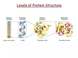
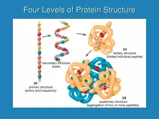
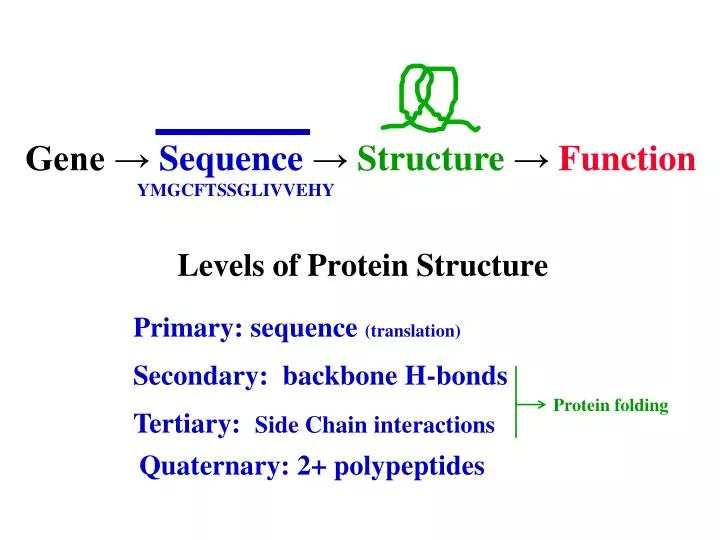
Levels of Protein Structure. Gene → Sequence → Structure → Function YMGCFTSSGLIVVEHY. Primary: sequence (translation). Secondary: backbone H-bonds. Protein folding. Tertiary: Side Chain interactions. Quaternary: 2+ polypeptides. Chromatography Types.

E N D
Levels of Protein Structure Gene →Sequence→Structure→ Function YMGCFTSSGLIVVEHY Primary: sequence (translation) Secondary: backbone H-bonds Protein folding Tertiary: Side Chain interactions Quaternary: 2+ polypeptides
Chromatography Types a) Size exclusion (gel filtration) b) Ion Exchange c) Affinity Which type of chromatography has pores in the solid matrix beads? Types of PAGE Which type of chromatography is most closely related to the function of the protein? a) Standard b) SDS c) Isoelectric focusing Which type of PAGE is used to determine the MW of a protein? Given the MW and pI data for a collection of proteins, which type of PAGE would be most difficult to predict?
Potato Extract Octyl-sepharose Stds 250k 150k 100k 75k 50k 37k 25k 20k 15k 10k DEAE G50 G50 ← PPO? ~68K ← STA ? ~60k ←Patatin→ ~ 43k ← WIN? ~19k If Catechol Oxidase is about 40k? It would migrate very close to patatin
Levels of Protein Structure Gene →Sequence→Structure→ Function YMGCFTSSGLIVVEHY Primary: sequence (translation) Secondary: backbone H-bonds Protein folding Tertiary: Side Chain interactions Quaternary: 2+ polypeptides
Sequence→Structure YMGCFTSSGLIVVEHY Protein Folding a spontaneous process in cells Unfolded & inactive folded and active
FREE ENERGYany spontaneous process must have DG < 0e.g. protein folding, celluar reaction, transport across membranes Enthalpy is minimized by forming bonds Entropy is maximized by increasing disorder G = H-S Melting/freezing Protein folding/unfolding ssDNA/dsDNA
equilibrium steady state non-equilibrium ↓ Level drops ↔ maintains level “A Cell is always striving to achieve a state of equilibrium, but never succeeding” “ An organism at equilibrium is a dead organism!”
Today’s topics: Levels of Protein Structure Secondary Structure – peptide bond planarity Fibrous Proteins - Collagen In order for a process to proceed which of the following must be true? a) DH > 0 b) DS > 0 c) DG < 0 d) DH < 0 As a protein folds into a functional structure which of the following is true? a) DH < 0 b) DS < 0 c) Both of the above d) neither of the above
Bonding Review G Minimize G DG is (-) covalent bonds (e.g. peptide bond) ionic bonds (salt bridges) F = q1q2/(r2D) hydrogen bonds (directional) dispersion forces (hydrophobic interactions) Why do proteins fold? G = H-S
O keto || — C — N — | H Planarity of Peptide Bond OC C N CH C N OH enol | — C = N — The backbone ‘direction’ can be defined by the F & Y angles for each Ca.
The polypeptide is made up of a series of planes linked at α carbons The backbone ‘direction’ can be defined by the F & Y angles for each Ca.
Secondary Structure (3 patterns) Due to the formation of H-bonds between the amide H and the carbonyl O atoms in the backbone of a polypeptide chain a-Helix ―coiled – every 4thaa is H-bonded b-Sheet ―extended - must have 2 or more segments b-Turn ―4 aa segment – reverses direction — C — || O
1.5å (max = 3.5å) a-Helix ―coiled – every 4thaa is H-bonded
a-Helix ―coiled – every 4thaa is H-bonded 1.5å ca to ca
a-helix in cranbin Side chains are too large to fit inside helix.
b-sheet is composed of 2 or more extended polypeptide segments from same chain. ← 3.5å ca to ca →
b-sheet strands are represented as arrows in protein backbone structure representations.
b - turn a - helix b - sheet Cranbin no formal secondary structure
The x-ray structure indicates F & Y angles. The F/Y angle pattern indicates the 2ndary structure.
a-helix b-sheet
b-sheet a-helix Ramachandran Plot (CPA) +180 -180 -180 +180
Myoglobin helices
HbRamachandran Plot some b-turn a-helix is dominant
What interaction creates protein secondary structure? a) covalent bonds b) ionic bonds/salt bridges c) H-bonds d) dispersion forces/hydrophobic interactions Which type of secondary structure has the longest distance between consecutive a-carbons? a) a-helix b) b-sheet c) b-turn d) 2ndary structure doesn’t influence peptide length
In a polypeptide backbone which type of bond cannot rotate? a) C=O to N b) N to Ca c) Cato C=O d) none of the above
Levels of Protein Structure Gene →Sequence→Structure→ Function YMGCFTSSGLIVVEHY Primary: sequence (translation) Secondary: backbone H-bonds Protein folding Tertiary: Side Chain interactions Quaternary: 2+ polypeptides
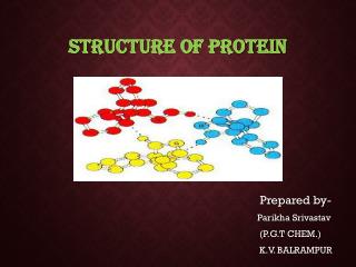
Types of Tertiary Interactions Disulfide ― A covalent bond between the S atom from two Cysteine side chains. H-bonds ― A hydrogen bond between any two polar (or acidic/basic) side chains Salt bridge ― An ionic bond between two oppositely charged side chains. One must be acidic and the other basic with the pH between the two pK values. Hydrophobic interactions ― Dispersion forces between a group of nonpolar side chains. Driven by the desire to avoid water causing ↑DS. Water is more ordered when its H-bonding options are limited by adjacent nonpolar groups.
TERTIARY STRUCTURE Disulfide Bond 2 Cys covalent C-S S-C H-bond 2 polar O-H O=C NH2 + Salt Bridge Acid/Base NH3O-C O
Nonpolar Groups Fold In to Avoid Water COO + H3N CH2 CH2 CH2 CH2 S CH2 CH3 CH2 CH3 CH3 CH-CH3 CH2 OH CH3 HO C=O NH2
insoluble cross-linked Structural function water soluble folded Fibrous vs. Globular Proteins
Fibrous Protein Examples: a-Keratins: hair, nails, skin - a-helical structure of chains b-Keratins: feathers, scales ... - b-sheet structure of chains Fibroin: silkworms, spider silk - b-sheet structure of chains Collagen: ~1/3 of animal protein skin, bones, connective tissue, teeth, gums .... etc
Amino Acids & Nutrition RDI = 60 g Gelatin is boiled collagen It contains few essential amino acids The sequence repeats Glycine (small) & Proline .... - Gly - xxx - Pro -... GPRGPAGPPGRDGIPGQPGLPGPPGPPGPPGPPGLGGNF
Collagen polypeptides form a triple helix structure.
GPRGPAGPPGRDGIPGQPGLPGPPGPPGPPGPPGLGGNF What is the role of glycine and proline in the function(structure) of collagen? Collagen Triple Helix Glycine’s small size is required for the tight weave of the three polypeptide chains into the triple helix? Proline allows for H-bonded cross-links to form within the triple helix and between strands, but only after it is modified to hydroxyproline.
N Ca C O CH2 CH2 CH2 OH Proline (Pro) Hydroxyproline (Hyp) How would you classify Proline as a side chain? a) nonpolar b) polar c) acidic d) basic Prolyl Hydroxylase How would you classify Hydroxyproline as a side chain? a) nonpolar b) polar c) acidic d) basic
Proline Hydroxylase (PH) - lumen of ER - Requires Fe and ascorbic acid (vitamin C). PH(Fe2+)+ O2 + a-ketoglutarate + Xaa-Pro-Gly PH catalyzes hydroxyproline formation PH(Fe2+)+ succinate + Xaa-Hyp-Gly Also …. PH(Fe 2+)+ O2 + a-ketoglutarate succinate + … PH(Fe3+)(not functional until reduced back to Fe2+) + Vit. C (red) PH(Fe2+)+ vit. C (ox)
Collagen Sequence About half the Pro residues are converted into Hyp (hydroxyproline). Gly-Pro-Met-Gly-Pro-Ser-Gly-Pro-Arg- Gly-Leu-Hyp-Gly-Pro-Hyp-Gly-Ala-Hyp- Gly-Pro-Gln-Gly-Phe-Gln-Gly-Pro-Hyp- Gly-Glu-Hyp-Gly-Glu-Hyp-Gly-Ala-Ser---- Hyp CH2 - CH2 - CH - OH
CH2 H O - CH - CH2 | CH2 - CH2 - CH - OH H-bonds cross-link collagen chains
Scurvey : Disease associated with Vitamin C deficiency ...caused by weakness in connective tissue ... caused by failure to form sufficient cross-links in collagen ... caused by insufficentvitamin C for ProlineHydroxylase (Fe3+) reduction Should you make sure your pet dog gets enough vitamin C? a) yes b) no Vitamin C is synthesized metabolically in all animals except …. humans, bats, guinea pigs, monkeys/apes (some fish/birds?)
OsteogenesisImperfecta Disease of? A) muscle b) nerves c) blood d) bones In what movie did Samuel L. Jackson have OI? Four types with varying severity ― I, II, III, IV Cause: mutation in gene for procollagena-1 chain Effect: interfere with 3-helix formation collagen used to make bone is weaker Procollagena-1 chain: mutations Gly221 Cys OI mild form Gly389 Cys OI moderate form Gly425 Ser OI lethal form Why do you think mutations of Glycine are causes of OI? a) Gly is small b) Gly forms H-bonds c) Gly is nonpolar
Soluble Collagen Disease SColD
$$ $$ Save The Witches Fund c/o Paul Stein Department of Chemistry The College of St. Scholastica
Hemoglobin (Hb) a2b2 4 subunits Quaternary structure ― The association of 2 or more folded polypeptides to form a functional protein molecule ― held together by H-bonds, salt bridges, hydrophobic interactions, and sometimes disulfide bonds.
WGA : Monomer Domains B C A D
Denaturing agents ― Cause proteins to unfold when added to solution in sufficient concentration. Side chains can form interactions with solute rather that other side chains.
Ribonuclease refolds spontaneously into its correct structure when urea removed. This requires a small amount of HO-CH2-CH2-SH to prevent incorrect disulfide bonds.