Download
1 / 32
540 likes | 3.08k Views
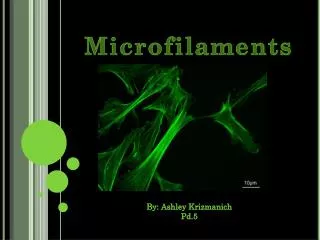
Microfilaments. Microtubules Microfilaments. Text and image sources are included using the notes function of this file. minus. Microfilament (thin filament) structure. G-actin monomer. ATP. --- 7-8nm. 25nm. plus. G-actin (42-43kD; 375 aas). (minus)

E N D
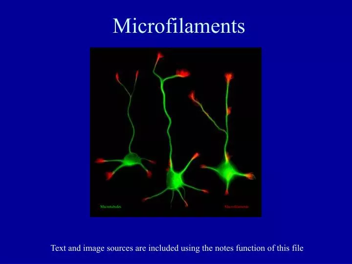
Microfilaments MicrotubulesMicrofilaments Text and image sources are included using the notes function of this file
minus Microfilament(thin filament) structure G-actin monomer ATP --- 7-8nm 25nm plus
G-actin (42-43kD; 375 aas) (minus) (plus) Changes upon hydrolysis Animation
Polarity of assembly(in vitro assay) Video + G-actin, ATP Red arrow = minus end Green arrow = initial plus end Bulls-eye = contact point on surface + -
+ S1 Decoration -
S1 decoration reveals microfilament polarities in situ a. Cytoplasm (mixed polarities) b. Stress fibers (bundled mixed polarities) c. Lamellipodia (uniform polarity, points in)
The Angel of Death Mushroom Amanita phalloides Phalloidin
Cells labeled with Phalloidin (F-actin) and DNase I (G-actin)
Actin Binding Proteins (ABPs) modulate architectures “. . . the actin cytoskeleton is a complex three-dimensional molecular jigsaw puzzle” - Tom Pollard
Profilin is involved in ATP recharge profilin
Cofilinweakens subunit interactionsby structuralintrogression
Actin subunit Amoeboid motility uses Gelsolin Gelsolin (partial structure) in presence of Ca++ Video
F-actinmotilities Composite time lapse segments
Actin dynamics at the leading edge GFP::actin in a tissue culture cell 2 time-lapse video sequences
Axonal growth cones Video GFP-actin dynamics Video Microtubules are excluded from the periphery Cell microinjected with fluorescent tubulin to label microtubules
Keratocyte motility Video
Arp2/3 immunolocalization Branching out on the family tree: the Arp2/3 complex plus 70o minus Nucleation Promoting Coronin Factors (NPFs) WASp/SCAR/Wave
Formin those long filaments - + G-actin monomer
WASp SCAR Assembly Cycles ATP translation Formin Arp2/3 VASp Dynamic F- actinADP microfilament CCT G-actinATP ADP (exchange) ATP PIP2 Profilin Stabilizing ABPs WASp VASp Thymosin Stable F-actin microfilament G-actinADP Severin Gelsolin
F-actin And VASp NPF: VASp(vasodilator stimulated phosphoprotein) Video
Signal transduction links G-proteins WASp WASp*P PIPkinase MLCK Cdc42 Rac Rho Filipodia Localized Initiation Lamellipodia Frontal Initiation Increased Turnover Stress fibers Form and Tension Arp2/3 PIP PIP2 Myosin Formin
Stress fibers distribute forces Arrows highlight microtubules adjacent to the stress fiber bundle
Neutrophil motility Video
Stereocilia MyoVIIa is involved in Ca++ regulated adaptation
Act’in Up Video Listeria monocytogenes moving in PtK2 cells