Download
1 / 48
480 likes | 498 Views
“THE GREAT MASQUERADER”. PULMONARY EMBOLISM. Dr. Prakash Mohanasundaram EMERGENCY PHYSICIAN. DEFINITION. Triad: Hypercoagulability Stasis to flow Vessel injury. HYPERCOAGULABILITY Malignancy Pregnancy Postpartum status(<4 wks) Estrogen Antiphospholipid antibodies

E N D
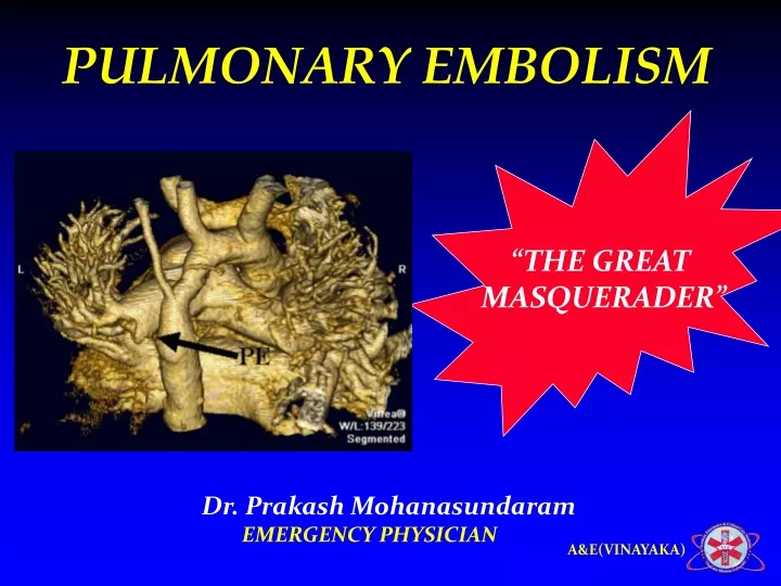
“THE GREAT MASQUERADER” PULMONARY EMBOLISM Dr. Prakash Mohanasundaram EMERGENCY PHYSICIAN
Triad: • Hypercoagulability • Stasis to flow • Vessel injury
HYPERCOAGULABILITY Malignancy Pregnancy Postpartum status(<4 wks) Estrogen Antiphospholipid antibodies Genetic mutations Factor V Leiden mutation Prothrombin gene mutation Factor VIII mutations Protein C deficiency Protein S deficiency VENOUS STASIS Bed rest >48 hrs Cast or external fixator Recent hospitalisation Long distance automobile or air travel RISK FACTORS • VESSEL INJURY • Recent surgery requiring endotracheal intubation • Recent trauma requiring hospitalisation
PATHOPHYSIOLOGY • Embolization • Physiology • Right ventricular dysfunction
EMBOLIZATION • Proximal leg DVT • Calf vein thrombi • Upper extremity thrombosis
PHYSIOLOGY • Increased pulmonary vascular resistance • Impaired gas exchange • Alveolar hyperventilation • Increased airway resistance • Decreased pulmonary compliance
DEATH “RIGHT VENTRICULAR DYSFUNCTION”
Clinical Features Symptoms in Patients with Angio Proven PTE Symptom Percent Dyspnea 84 Chest Pain, pleuritic 74 Anxiety 59 Cough 53 Hemoptysis 30 Sweating 27 Chest Pain, nonpleuritic 14 Syncope 13
Clinical Features Signs with Angiographically Proven PE Sign Percent Tachypnea > 20/min 92 Rales 58 Accentuated S2 53 Tachycardia >100/min 44 Fever > 37.8 43 Diaphoresis 36 S3 or S4 gallop 34 Thrombophebitis 32 Lower extremity edema 24
NON IMAGING D-Dimer ELISA ABG ECG NON INVASIVE CXR Venous ultrasonography Chest CT Lung scanning MR Contrast enhanced Echocardiography DIAGNOSING MODALITIES INVASIVE • Pulmonary angiography • (GOLD STANDARD) • Contrast phlebography
D-dimer Test • Fibrin split product • Circulating half-life of 4-6 hours • Positive assay > 500 ng/ml • Quantitative test have 80-85% sensitivity, and 93-100% negative predictive value • False Positives: Pregnant Patients Post-partum < 1 week Malignancy Surgery within 1 week Advanced age > 80 years Sepsis Hemmorrhage CVA AMI Collagen Vascular Diseases Hepatic Impairment
ABG • Hypoxemia • Hypocarbia “ LACK DIAGNOSTIC UTILITY IN PE ”
ECG • Most Common Findings: • Tachycardia or nonspecific ST/T-wave changes • Acute cor pulmonale or right strain patterns • Tall peaked T-waves in lead II (P pulmonale) • Right axis deviation • RBBB • S1-Q3-T3 (occurs in only 20% of PE patients) • Atrial fibrillation / Atrial flutter
Chest X ray • Westermark’s sign focal oligemia / cut off sign • Hampton’s hump peripheral wedge shaped density above the diaphragm • Palla’s sign enlarged right descending pulmonary artery ALMOST ALWAYS NORMAL CHEST X RAY
Venous Ultrasonography Loss of vein compressibility ½ of pts with PE have no imaging evidence of DVT
Chest CT • Principal imaging test • New generation multislice scanners locates thrombi in the fifth order branches • Alternative diagnosis • Pneumonia • Emphysema • Pulmonary fibrosis • Pulmonary mass • Aortic pathology
MR contrast enhanced • Results similar compared with first generation CT • Also assesses right ventricular function
Echocardiography • ½ pts have normal echo • DD’s • AMI • Pericardial tamponade • Aortic dissection • PE complicated by right heart failure • Risk stratification MC CONNEL’s sign – right ventricular free wall hypokinesis with normal right ventricular apical motion
Pulmonary angiography(GOLD STANDARD) • Detect emboli as small as 1 to 2 mm RESERVED FOR • Technically inadequate CT scans • Scans performed on older machines • Pts who will undergo interventions
Arrow indicates abrupt termination of a pulmonary artery. Www.brighamrad.Harvard.edu/cases/bwh/images. Pulmonary Embolus
TREATMENT THE EMERGENCY PERSPECTIVE
DICTUM “ABC”
PRIMARY THERAPY Thrombolysis Embolectomy ADJUNCTIVE THERAPY O2 Pain relief Dobutamine Caution – volume overload TREATMENT SECONDARY THERAPY • Anticoagulation • IVC filters Pulmonary thromboendarterctomy
SCENARIO • 45 year male, case of OPC poisoning • Being treated with mechanical ventilation • Paralysed & sedated for 2 days • Develops sudden tachypnoea, tachycardia, hypotension & hypoxia
THROMBOLYSIS • Recombinant tPA 100 mg iv infusion over 2 hours • Streptokinase 250,000 U iv over 30 mins foll by 100,000 U/hr for 24 hrs • Urokinase 4,4OO U/kg iv over 10 mins foll by 4,000 U/kg/hr for 12 hrs • Alteplase 15 mg iv bolus foll by 2 hr infusion of 85 mg ( discontinue heparin during infusion)
SCENARIO • 45 year male, A case of glioma • Underwent craniotomy & evacuation 2 days ago • Bed ridden for 2 days • Develops sudden tachypnoea, tachycardia, hypotension & hypoxia
EMBOLECTOMY Indicated in pts with risk of thrombolysis • Surgical embolectomy • Catheter embolectomy
SCENARIO • 45 year male, case of OPC poisoning • Being treated with mechanical ventilation • Paralysed & sedated for 2 days • Develops sudden tachypnoea & tachycardia • BP - Normal
ECHO NORMAL WHAT IS YOUR LINE OF MANAGEMENT
Heparin / LMWH / Warfarin • Heparin 80 U/kg iv bolus foll by 18 U/kg/hr • Enoxaparin 1 mg/kg twice daily / 1.5 mg/kg daily • Tinzaparin 175 mg/kg OD • Fondaparinux <50 kg receive 5 mg, 50–100 kg patients receive 7.5 mg >100 kg receive 10 mg. • Warfarin – 2.5 to 10 mg Target INR – 2.0 TO 3.0
IVC Filters • INDICATIONS • Active bleeding that precludes anticoagulation • Recurrent venous thrombosis despite intensive anticoagulation
SUMMARY • > 50 % pts with DVT are associated with PE • > 50 % cases do not have any signs or symptoms • Common presentation can be unexplained tachycardia, tachypnoea, hypoxemia or mere anxiety • Diagnosis and suspicion is purely clinical • Follow up with anticoagulants is must as there is a increased risk of recurrence
PREVENTION IS BETTER THAN CURE