Download
1 / 19
220 likes | 829 Views
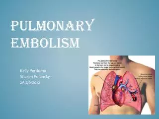
Pulmonary Embolism. Radiologic Evaluation. Lucas Faulkenberry M4. Objectives. Understand radiologic studies used to identify pulmonary embolism Identify abnormalities in these studies Review the new algorithm for evaluating suspected PE. Pulmonary Embolism. Non-specific symptoms CP SOB

E N D
Pulmonary Embolism Radiologic Evaluation Lucas Faulkenberry M4
Objectives • Understand radiologic studies used to identify pulmonary embolism • Identify abnormalities in these studies • Review the new algorithm for evaluating suspected PE
Pulmonary Embolism • Non-specific symptoms • CP • SOB • Tachycardia • Tachypnea • Cough • Hemoptysis • Frequent autopsy finding • 80% associated with DVT
Diagnostic Efforts in Radiology • reaching an acceptable level of diagnostic certainty of PE to warrant anticoagulant therapy, using the least invasive tests • eliminating other reasons for the patient's symptoms
Acute Chest Pain with Suspected PE • Leg doppler • CXR • V/Q Scan • CTPA • Pulmonary Angiography
Leg Doppler • Quick • Cheap • Non-invasive • Positive test leads to same treatment
Chest X-Ray • Important initially to rule out obvious causes of CP • Abnormal in 88% • Hampton’s Hump • Westermark Sign
V/Q Scan • Not as widely used • Allergy or pregnancy • PIOPED criteria
Computed Tomography Pulmonary Angiography • New standard for suspected PE • Fast • High sensitivity and specificity • Few contraindications
Pulmonary Angiography • Used less often • Confirmation or indeterminate cases • Invasive
Review • Imaging studies used to evaluate suspected PE • Practice identifying abnormalities in these studies • Review the new algorithm for evaluating suspected PE