Download
1 / 40
400 likes | 402 Views
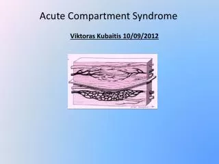
Herniation: Compartment Syndrome of the Head. Connie Chen, MD Neurology Consultants of Dallas. Goals. Understand Recognize Treat. Theory. Compartment = skull contents Fixed volume: Brain (80%), blood (10%), csf (10%) Stable pressure: CPP=MAP-ICP. Theory.

E N D
Herniation:Compartment Syndrome of the Head Connie Chen, MD Neurology Consultants of Dallas
Goals • Understand • Recognize • Treat
Theory • Compartment = skull contents • Fixed volume: • Brain (80%), blood (10%), csf (10%) • Stable pressure: CPP=MAP-ICP
Theory • Autoregulation of increased intracranial pressure (cough/valsalva): Increased ICP Increase MAP Compartment’s goal: Maintain CPP!!
Another look • Perfusion of a compartment: CPP= MAP- ICP If MAP cannot increase: • Increased ICP = Decreased CPP • Decreased CPP = Tissue ischemia • Tissue ischemia = Edema
Another look • Edema = Further increased ICP • Further decreased CPP = Tissue death
What Else? • Monro-Kellie doctrine: - Skull is a fixed volume - increase in one volume leads to decrease in others. Brain> blood, csf
What Else? Increased ICP (via increased volume) Displace blood/csf **Displace brain** !!!!
Theory Increased ICP via increased volume Displaces blood, CSF, then brain & Reduces CPP causing brain ischemia
Recognize Looking at a Head CT is not recognition
Recognize Displaced brain will cause neurologic signs
Signs falx • Where does displaced brain go? • Side to side: subfalcine • Side to bottom: uncal (transtentorial) • Top to bottom: central tentorial • Bottom to top: “upward” • Bottom thru the “hole”: tonsillar tentorium foramen magnum
Signs • Subfalcine • ACA compression: contralateral leg paresis • Somnolence • Uncal (transtentorial) • Anisocoria to “blown pupil” • Midbrain and PCA compression: Somnolence, Contralateral hemiparesis, occipital infarct • Decerebrate posturing (extensor) midbrain
Signs • Central tentorial • Somnolence/coma • Bilaterally “blown” pupils • Decorticate/decerebrate posturing • Bilateral midbrain, PCA compression • Upward (rare) • Midbrain compression • “Blown” pupils • Somnolence/coma midbrain
Signs • Tonsillar • Somnolence • Quadriparesis • Cardiac arrythmias • Respiratory failure medulla midbrain Foramen magnum
More Signs • Vital sign changes (brainstem is being crushed): “Cushing Reflex” : Bradycardia/hypertension respiratory change • Somnolence/Coma • EXAMINE PT: pupils, pupils, pupils
Pupils? • Blown pupils: (large unreactive) • Not medication unless ophthalmology came • Or if under GENERAL anesthesia • Compression of midbrain • Pinpoint pupils: (small unreactive) • Often caused by medication (benzo’s, opiates) • Also from pontine damage
Pupils? • Anisocoria: • Sometimes normal or surgical • Sometimes meds: nebulizer • Compression of CN III • Irregular: • Surgical • Ongoing ischemia: “cat eye”
Exam Summary • Vital sign change is LATE • Early exam change- somnolence • Pupil change- anisocoria or less reactive • Very late change: comatose, dilated pupils, posturing
Treatment Head CT is not a treatment
Treatment • Herniation = Brain CODE • Stabilize your pt first: ABC’s, then BCB • Then conduct “secondary survey”
Treatment • Control your compartment (BCB) • Blood • CSF • Brain
Blood • Allow outflow: • Cut the tape off of the neck • Head midline • Head of bed at 45 degrees • Constrict blood vessels: • Hyperventilate: BAG! • Goal is pH change- not pCO2
CSF • Drain CSF • Intraventricular catheter placement • Spinal CSF removal will let you herniate faster
Brain Steroids only work on tumors
Brain • Shrink • Hyperosmolar agents: mannitol, hypertonic saline • Doses? Bolus vs infusion. • Cut • Surgical removal • Shut down • Neuroanesthetic agents: propofol, thiopental
How Long? Hyperosmolar agents and hyperventilation lasts 6 hours at best.
Then what? • Your pt is better • Your pt is not better
Then what? Head CT verifies your diagnosis and identifies the problem
Release • Release the compartment : “pop the top” • Early vs. late
Case 60 yo wm s/p acute right MCA stroke
Case • 4 days later • Pt becomes somnolent but arousable • There’s new anisocoria • Plan?
Case • 12 hours later • Not arousable- comatose • Still anisocoria, right pupil stops reacting • Plan?
Case Other options?
Summary • Look outside your “box” • Herniation is reversible • Treat before scanning