Download

1 / 19
240 likes | 614 Views
Foot compartment syndrome. Ryan Thomas. Description. The traumatic event usually causes an initial increase in the interstitial fluid Decreased capillary blood flow and local muscle ischemia occur gradually. This ischemic process promotes vasodilation and increased capillary permeability.

E N D
Foot compartment syndrome Ryan Thomas
Description • The traumatic event usually causes an initial increase in the interstitial fluid • Decreased capillary blood flow and local muscle ischemia occur gradually. • This ischemic process promotes vasodilation and increased capillary permeability. • Additional intracompartmental edema and increased tissue pressure. • Ischemic muscle undergoes necrosis, fibrosis, and contracture. • Nerves can sustain compression for longer periods than muscles and show some reversibility, particularly as it relates to sensation.
Causes • High velocity injuries, multiple fractures, crushing injuries, dislocations, calcaneal fx, lisfranc’s fx, blood dyscrasias, arterial injury/embolism, sepsis/shock, surgery (artery clamping), repetitive/excessive muscle use (exercise with dehydration, tetanus, seizures, delirium tremens), cast, tourniquet.
Manoli and Weber postulated that the claw toe deformity, following a calcaneus fracture, appears to be secondary to a late contracture of the quadratus plantae muscle in the calcaneal compartment. • Myerson and Manoli postulated that the pathologic process proceeds as a result of the large bleeding cancellous bone surfaces. Calcaneal compartment pressures rise as the hematoma attempts to dissipate into limited osseofascial compartments. • As the pressure rises, the quadratus plantae muscle becomes ischemic within the calcaneal compartment.
Myerson retrospectively looked at 14 compartment syndromes in 12 patients. Of the crush injuries, 41% developed compartment syndrome compared with 17% of noncrush injuries (calcaneal fractures). The initial energy expanded at the time of trauma plays a crucial role in determining the timing and development of FCS. • can develop 36 hours after the inciting event
Signs • The clinical signs of FCS are vague and ill defined compared with the classic presentations of compartment syndrome of the lower extremity. Patients with calcaneal fractures and subsequently proven FCS describe clinical symptoms of a severe, relentless, burning pain involving the entire foot.
Signs • Neurological signs such as paresthesia, anesthesia, and pain begin after only a short ischemic period (1-1.5 hours) • Muscle weakness is evident at 3-4 hours • Irreversible muscle tissue damage starts at 4-6 hours • 6 Ps – pain, pressure, paresthesia, paresis, pain with passive stretch, and pulselessness • Pulselessness is an unreliable sign since pressures need not be suprasystolic to produce compartment syndrome
Pressures • Normal pressures in foot are 5 +/- 3 mmHg with a range of 1-12 mmHg • Generally pressures of 0-20 mmHg is considered normal • Pressures of 20-30 mmHg are borderline • Pressures in the range of 30-45 mmHg in the face of clinically suggestive symptoms should be treated with a fasciotomy • A pressure of 45 mmHg alone is diagnostic of compartment syndrome and mandates a fasciotomy
Findings • Vibratory, 2 point, and light touch are better than pin prick test. • presence of tense swelling • only way of diagnosing compartment syndrome reliably is direct tissue pressure measurement. • Pain, sensory, and motor changes often are not as dramatic in the foot as in other locations that develop a compartment syndrome. • transcutaneous oximetry may be helpful in assessing the adequacy of compartment decompression.
Muscle undergoes necrosis within 4 hours of ischemia • Fibroblasts replace the infarcted muscle. This process can progress over 6 to 12 months. Additionally, this necrotic muscle often adheres to surrounding tissues, which fix the muscle position and reduce mobility further. • It is thought that the limited muscle excursion as well as the longitudinal contraction during fibrotic proliferation results in the loss of joint motion and subsequent contracture.
Tx • All bandages, casts, and splints are widely opened or removed • Extremity is placed at heart level • Foot is monitored both clinically and with compartment pressures • Emergency fasciotomy should be within first 6 hours
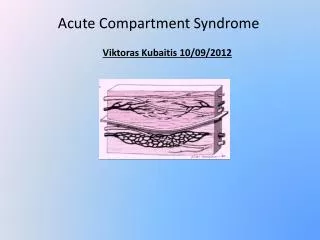
Foot compartment anatomy • 3 run entire length of the foot; medial, lateral, superficial. • 6 confined to forefoot or hindfoot; adductor, the four interosseoi, calcaneal • There is communication between calcaneal compartment and deep posterior compartment of the leg through flexor retinaculum.
Fasciotomy • Manoli and Weber recommend a medial incision for decompressing the medial compartment and reflecting the abductor hallucis muscle superiorly. • Next the fibrous intermuscular septum is opened longitudinally to release the calcaneal compartment. The superficial compartment is released by following the medial surface of the medial compartment, which decompresses the flexor digitorum brevis. This muscle is retracted plantarward, which allows access to the lateral compartment containing the abductor digiti minimi and flexor digiti minimi brevis. Two dorsal incisions are used to decompress the individual interosseous compartments. The adductor hallucis compartment is approached by stripping muscles off the medial aspect of the second metatarsal. • Patients who had undergone fasciotomy typically underwent skin closure 5 to 10 days after the procedure.
if the recognition of compartment syndrome is delayed for more than 8 to 10 hours, the traditional operative treatment of fasciotomy should be reassessed. • If a patient presents later to the office with a history compatible with the development of FCS and development of contractures consistent with the sequelae of compartment syndrome, an aggressive rehabilitation program is started immediately.
Tx for complications • Operative management is reserved for the treatment of residual nerve compression and problematic deformities. Surgical options include (1) release of fixed contracture, myotendinous lengthening, muscle resection, or tenotomy; (2) tendon transfers or fusion to increase function; and (3) ostectomy or amputation for severe refractory deformities. If the contracture is limited to one or two muscles and a cordlike area of induration is palpable, lengthening or release can be performed. If, the deformity is flexible and a reasonable donor muscle exists, a tendon transfer can be considered. Severe involvement requires an osteotomy, arthrodesis, or amputation. • Symptomatic claw toes often are treated surgically with conventional methods using a proximal interphalangeal resection at the level of the condylar heads with a flexor to extensor tendon transfer. All symptomatic claw toes are treated, not just the second and third toes. After surgery, the toe should be straight and passively flexible with a normal appearance. • Nerve decompression alone rarely is indicated; however, posttraumatic scarring of the nerve occasionally requires external neurolysis. An additional source of nerve irritation is fibrosis of the contiguous muscle and tendon with scar formation.
References • Manoli A II, Weber TG: Fasciotomy of the foot: An anatomical study with special reference to release of the calcaneal compartment. Foot Ankle 10:267-275, 1990 • Orthop Clin North Am. 2001 Jan;32(1):103-11.Foot compartment syndrome.Perry MD, Manoli A 2nd.Department of Orthopedic Surgery, University of South Alabama College of Medicine, Mobile 36617, USA.