Download

1 / 26
280 likes | 542 Views
Folding and flexibility. Outline. What is protein folding ? How proteins fold in vivo ? What is protein flexibility ?. Structural features present in any folded globular protein:. 1. Most mainchain hydrogen bonds are formed.

E N D
Outline • What is protein folding ? • How proteins fold in vivo ? • What is protein flexibility ?
Structural features present in any folded globular protein: • 1. Most mainchain hydrogen bonds are formed. • 2. Core is formed of hydrophobic residues, but not all hydrophobic residues are buried. • 3. Well-packed hydrophobic core. • 4. Secondary structural elements (helices, sheet) show a hydrophobic and hydrophilic face - Amhipathic helices have hydrophobic residues every third/fourth residue - Amphipathic sheets alternate hydrophobic and hydrophilic residues. • 5. Most polar residues are on the surface. • 6. Buried polar residues have hydrogen bonded partners. • 7. Almost all charged residues are on the surface. • 8. Buried charged residues often have charges of opposite sign close by.
Properties of molten globule • Most secondary structure elements of native state have formed • Less compact than native state and proper packing of hydrophobic core is not yet accomplished • Interior is in more “liquid” than solid state • Surface structures and loops largely unfolded
How do correct disulfides form ? • Example: Bovine pancreatic trypsin inhibitor is unfolded before the disulfides have formed • Protein disulfide isomerase catalyzes internal disulfide exchange • During folding, first disulfides form in random • More stable disulfides accumulate
Prolyl peptide isomerase • Active site is on the side of barrel, which is quite unusual • The cis-trans isomerization is increased 1,000,000-fold
Molecular chaperones • Proteins, which help the other proteins to fold corrcetly • How?
Chaperonin GroES-GroEL • GroEL : 14 subunits • GroES : 7 subunits
Structure of GroEL monomer Unfolded proteins bind to hydrophobic residues of apical domain ATP binds to equatorial domain
Binding of GroES and ATP to GroEL • Upon binding of GroES and ATP, one side of GroEL gets extended and the central cavity increases in volume • In central cavity now are less hydrophobic residues exposed • The other side of GroEL looses affinity to another GroES molecule
Events upon protein folding inside GroES-GroEL complex unfolded protein folded protein
Chaperones, other than GroES-EL exist • Principle is similar – chaperones bind to hydrophobic surfaces • Other chaperones do not form enclosed structure
Can the 3D structure of aprotein be predicted? • Many proteins can be folded andunfolded reversibly in vitro • This implies that practically allinformation necessary to determinethe 3D structure is contained in thesequence
But... • A general method for accurate fold prediction has not been discovered yet • Available methods are at most 60% reliable and certainly not a replacement for x-ray or NMR studies
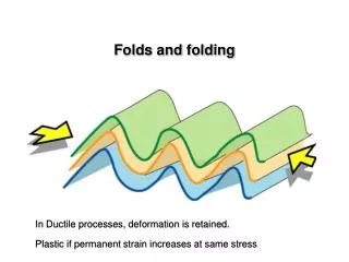
Flexibility • Folded proteins are not static – structural rearrangements are common • In most cases the structural rearrangements are minor and limited to loop regions • Frequently, some particular secondary structure element (strand or helix) switches between ordered / disordered states (for example, upon ligand binding) • Sometimes, major structural rearrangements are found
Causes of structural rearrangemenets • Interaction with a ligand • Interaction with other proteins • Changes of pH and/or ionic strength • Covalent modifications
Examples of minor structural rearrangements • Different packing environments for icosahedral virus MS2 coat protein subunits Same loop adopts different conformations at vertices of icosahedron and in the middle of one triangular face
Ordered C-terminal helix upon substrate binding to glutathione transferase
Example of major structural rearrangements • Conformational changes of calmodulin
Flexibility in multidomain proteins • Example: fibonectin • Large extracellular protein with several functions • Composed from about 30 domains • All linker regions are flexible