Download

1 / 43
430 likes | 504 Views
Synapses and Synaptic Transmission Dr. Eman El Eter Physiology Dep. College of Medicine KSU. Objectives. Define synapses Functions of synapses. Structure of synapses Types of synapses: anatomical & functional. Synaptic transmission & neurotransmitters Fate of neurotransmitters.

E N D
Synapses and Synaptic Transmission Dr. Eman El Eter Physiology Dep. College of Medicine KSU
Objectives • Define synapses • Functions of synapses. • Structure of synapses • Types of synapses: anatomical & functional. • Synaptic transmission & neurotransmitters • Fate of neurotransmitters. • Electrical events at synapses (EPSPs & IPSPs). • Properties of synaptic transmission • Factors affecting synaptic transmission
INTRODUCTION TO SYNAPSE: The CNS contains more than 100 billion neurons. Incoming signals enter the neuron through synapses located mostly on the neuronal dendrites, but also on the cell body. For different types of neurons, there may be only a few hundred or as many as 200,000 such synaptic connections from input fibers. Conversely, the output signal travels by way of a single axon leaving the neuron.
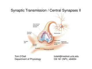
What is a synapse? A junction where the axon or some other portion of one cell (= presynaptic cell) terminates on the dendrites, soma, or axon of another neuron (post synaptic cell). The term was introduced in nineteenth century by the British neurophysiologist Charles Sherrington
Anatomical Types of Synapses • Axodendritic – synapses between the axon of one neuron and the dendrite of another • Axosomatic – synapses between the axon of one neuron and the soma of another • Other types of synapses include: • Axoaxonic (axon to axon) • Dendrodendritic (dendrite to dendrite) • Dendrosomatic (dendrites to soma)
Anatomical Types of Synapses Figure 11.17
Functional types of synapses A. Chemical synapse Almost all synapses used for signal transmission in the CNS of human being are chemical synapses. i.e. first neuron secretes a chemical substance called neurotransmitter at the synapse to act on receptor on the next neuron to excite it, inhibit or modify its sensitivity.
B. Electrical Synapses Membranes of the pre- and post-synaptic neurons come close together and gap junctions forms low membrane borders which allow passage of ions. • Are less common than chemical synapses • Correspond to gap junctions found in other cell types • Are important in the CNS in: Arousal from sleep • Mental attention • Emotions and memory • Ion and water homeostasis
C. Conjoint synapse Both electrical and chemical. Examples neurons in lateral vestibular nucleus.
What happens at the synapse? Information is transmitted in the CNS mainly in the form of APs “=nerve impulse”, which pass from one neuron to another. Each impulse through its way from one neuron to another may be:- • blocked in its transmission from one neuron to another • changed from single impulse to repetitive impulses. • Synaptic transmission is a complex process that permits grading and adjustment of neural activity necessary for normal function.
Examples of synapses outside CNS • NMJ • Contact between: autonomic neurons and smooth , cardiac muscles, and other effector cells.
SYNAPSE: STRUCTURE & FUNCTIONS Synaptic cleft: This the space between the axon terminal and sarcolemma. It has a width of 200-300 angstroms. Synaptic knobs (presynaptic terminal ) cover about 40% of soma and 70% of dendritic membrane
Action of the transmitter substance on post-synaptic neuron: At the synapse, the membrane of post-synaptic neuron contains large number of receptor proteins. Binding of the neurotransmitter to its receptor will result in inhibition or excitation of the post-synaptic membrane depending on the type of the neurotransmitter i.e. excitatory or inhibitory.
These receptors have two components 1. Binding site that face the cleft to bind the neurotransmitter 2. Ionophore: It passes all the way through the membrane to the interior. It is of two types 2nd messenger system in the post-synaptic membrane. This mechanism is important where prolonged post-synaptic changes are needed to stay for days, months . . Years (memory). Effects: intracellular enzymes activation, gene transcription, etc… Ion channels Cation channels Na+ (most common) K+ Ca++ Opening of Na+ channels membrane potential in positive direction toward threshold level of excitation (+) neuron Anion channels Cl¯ (mainly) Opening of Cl¯ channels diffusion of negative charges into the membrane membrane potential making it more negative away from threshold level (-) neuron
Fate of a neurotransmitter After a transmitter substance is released at a synapse, it must be removed by:- • Diffusion out of synaptic cleft into surrounding fluid • Enzymatic destruction e.g. Ach esterase for Ach • Active transport back into pre-synaptic terminal itself e.g. norepinephrine
Electrical events in post-synaptic neurons: • 1. RMP of neuronal soma: • ~65mV i.e. less than sk. ms. [70 to 90mV] • If the voltage is less negative the neuron is excitable • Causes of RMP: • Leakage of K+ (high K+ permeability) • Large number of negative ions inside: proteins, phosphate • Excess pumping of Na+ out by Na+-K+ pump
2. Effect of synaptic excitation on post-synaptic membrane: = Excitatory post-synaptic potential [EPSPs] When excitatory neurotransmitter binds to its receptor on post-synaptic membrane partial depolarization [ Na influx] of post-synaptic cell membrane immediately under presynaptic ending, i.e. EPSPs If this potential rises enough to threshold level AP will develop and excite the neuron ( via central or neuronal summation)
This summation will cause the membrane potential to increase from 65mV to 45mV. EPSPs = +20mV which makes the membrane reach the firing level AP develops at axon hillock. N.B. Discharge of a singlepre-synaptic terminal can neverincrease the neuronal potential from 65mV to 45mV.
EPSPs are characterized by: • Graded, unpropagated response. • Proportionate to the strength of the stimulus • Can be summated • If large enough to reach firing level AP is produced • Post-synaptic potential of +10 to +20mV is needed to produce AP..
3. Inhibitory post-synaptic potentials (IPSPs): • When an inhibitory neurotransmitter binds to its receptor on post-synaptic membrane, it causes hyperpolarization of the post-synaptic memb. which is the IPSP. • Causes: • An increase in membrane permeability toCl¯of post-synaptic memb. (produced by inhibitory neurotransmitter) excitability and memb. potential becomes away from firing level. • Also IPSP can be produced by:- • Opening of K+channels outward movement of K+ • Closure of Na+orCa++ channels • -IPSP = 5mV
Synaptic properties 1. One-way conduction Synapses generally permit conduction of impulses in one-way i.e. from pre-synaptic to post-synaptic neuron.
Properties of synapses (con…) 2. Synaptic delay Is the minimum time required for transmission across the synapse. This time is taken by • Discharge of transmitter substance by pre-synaptic terminal • Diffusion of transmitter to post-synaptic membrane • Action of transmitter on its receptor • Action of transmitter to membrane permeability • Increased diffusion of Na+ to post-synaptic potential
Properties of synapses (con…) 3. Synaptic inhibition Types: A. Direct inhibition B. Indirect inhibition C. Reciprocal inhibition D. Inhibitory interneuron
Properties of synapses (con…) A. Direct inhibition Occurs when an inhibitory neuron (releasing inhibitory substance) acts on a post-synaptic neuron leading to its hyperpolarization due to opening of Cl¯ [IPSPs] and/or K+ channels. Example : Glycine at the level of the spinal cord to block pain impulses.
Properties of synapses (con…) B.Indirect inhibition (=Pre-synaptic inhibition). This happens when an inhibitory synaptic knob lie directly on the termination of a pre-synaptic excitatory fiber. The inhibitory synaptic knob release a transmitter which inhibits the release of excitatory transmitter from the pre-synaptic fiber. The transmitter released at the inhibitory knob isGABA. The inhibition is produced by Cl¯ and K+. e.g. occurs in dorsal horn pain gating.
Properties of synapses (con…) C. Reciprocal inhibition Inhibition of antagonist activity is initiated in the spindle in the agonist muscle. Impulses pass directly to the motor neurons supplying the same muscle and via branches to inhibitory interneurones that end on motor neurones of antagonist muscle.
Properties of synapses (con…) D. Inhibitory interneuron ( Renshaw cells) Negative feedback inhibitory interneuron of a spinal motor neuron .
Properties of synapses (con…) 4. Summation • a.Spatial summation. • When EPSP occurs in more than one synaptic knob atthesame time. • b. Temporal summation. • If EPSPs in a pre-synaptic knob are successivelyrepeated without significant delay so the effect of the previous stimulus is summated to the next.
Properties of synapses (con…) 5. Convergence and divergence Convergence When many pre-synaptic neurons converge on any single post-synaptic neuron. Divergence Axons of pre-synaptic neurons divide into many branches that diverge to end on many post-synaptic neurons.
Properties of synapses (con…) 6.Occlusion • It is a decrease in the expected response due to pre-synaptic fibers sharing post-synaptic neuron [=overlap].
Properties of synapses (con…) 7. Fatigue It is due to exhaustion of neurotransmitter. If the pre synaptic neurons are continuously stimulated there may be an exhaustion of the neurotransmitter. Resulting in stoppage of synaptic transmission.
Factors affecting synaptic transmission: Alkalosis: Normally, alkalosis greatly increases neuronal excitability. For instance, a rise in arterial blood pH from the 7.4 norm to 7.8 to 8.0 often causes cerebral epileptic seizures because of increased excitability of some or all of the cerebral neurons. This can be demonstrated by asking a person who is predisposed to epileptic seizures to over breathe. The over breathing blows off carbon dioxide and therefore elevates the pH of the blood momentarily
Acidosis: Conversely, acidosis greatly depresses neuronal activity; A fall in pH from 7.4 to below 7.0 usually causes a comatose state. For instance, in very severe diabetic or uremic acidosis, coma virtually always develops.
Drugs: Many drugs are known to increase the excitability of neurons, and others are known to decrease excitability. Caffeine found in coffee, tea, increases neuronal excitability, by reducing the threshold for excitation of neurons.
Strychnine: Is one of the best known of all agents that increase excitability of neurons. It inhibits the action of some normally inhibitory transmitter substances, especially glycine in the spinal cord. Therefore, the effects of the excitatory transmitters become overwhelming, and the neurons become so excited that they go into rapidly repetitive discharge, resulting in severe tonic muscle spasms.
Hypoxia • Depression of neurons