Download
1 / 24
290 likes | 552 Views
Physiology of Synapses. Dr Taha Sadig Ahmed Physiology Department , College of Medicine , King Saud University , Riyadh. Objectives At the end of this lecture the student should : (1) define synapses and show where they are located .

E N D
Physiology of Synapses Dr TahaSadig Ahmed Physiology Department , College of Medicine , King Saud University , Riyadh
Objectives • At the end of this lecture the student should : • (1) define synapses and show where they are located . • (2) describe the parts of a synapse , & what does each part contain . • (3) know how to classify synapses . • (4) define synaptic transmitters , give examples of excitatory & inhibitory ones ; explain how they are released • (5) explain ionic channels that mediate actions on synaptic receptors . • (6) explain : EPSP , IPSP , LTP . • (7) describe properties of synapses such as convergence , divergence , spatial & temporal sunmmation , subliminal fringe , types of inhibition and their physiological significance . • (8) expalin how acidosis and alkalosis can affect synaptic transmission . References :Ganong Review of Medical physiology, 23rd edition . Barret et al ( eds) . Mc Graw Hill , Boston 2010 . Page 115 onward
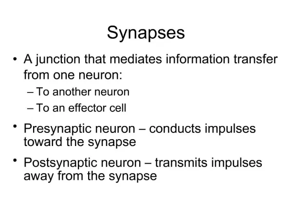
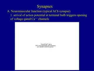
What is a synapse ? It is a n area of communication between 2 neurons . • What are its components & their function ? does each part of synapse contain ?
Components of a Synapse Q: What are the components of a synapse ? • Synaptic knob of the pre-synaptic cell ( contains transmitter ) (2) Synaptic cleft (space ) contains enzyme that destroys the transmitter (3) Post-synaptic membrane ( contains receptors for the transmitter )
Classification of Synapses (1) Axo-dendritic , ( 2) Axo-somatic , (3) Axo-axonicc , & less commonly (4) Dendro-somatic (5) Somato-somatic
Q : What are the types of transmitters ? • Excitatory neurotransmitter : a transmitter that produces excitatory postsynaptic potential ( EPSP) on the postsynaptic neuron . • Inhibitory neurotransmitter : a transmitter that produces inhibitory postsynaptic potential ( IPSP ) on the postsynaptic neuron .
Q : What are EPSP and IPSP ? • A : They are local responses • Q : What is their bioelectric nature ? • A : Graded Potentials ( i.e., proportional to the strength of the stimulus ). • Q: In what way do they affect the excitability of the postsynaptic membrane ? • A: EPSP makes the postsynaptic membrane more excitable ( thus more liable to fire AP ; & IPSP makes it less excitable) Q: In what ways do they differ from action potentials ? • (1) They are proportional to the strength of the stimulus ( i.e., do not obey All-or-None Law) • (2) They can summate ( add up )
Q : Give examples of excitatory transmitters ? • (1) Acetylcholine : Opens sodium channels in the Postsynaptic Cell Membrane depolarization EPSP . • (2) Glutamate : Produces EPSP by opening of calcium channels . • Q : What is long-term-potentiation ( LTP ) ?, what transmitter is involved in it ? What is the physiological function of LTP ?
Give examples of Inhibitory Tran smitters • When the inhibitory transmitter combines to its receptors , it produce Inhibitory Postsynaptic potential (IPSP) that hyperpolarizes the post-synaptic cell , thereby making it less excitable (more difficult to produce APs ) . • Examples of inhibitory transmitter is • GABA which in some places opens chloride channels , and in others opens potassium channels • Enkephalin Inhibitory transmitter . Found in the GIT and spinal cord . It exerts analgesic activity, reducing the feeling of pain . • Glycine ( mainly in spinal cord ) .
Formation of a Transmitter • Q : In what location of the neuron is the neurotransmitter synthesized ? • Q : In what location of the neuron is the transmitter vesicle synthesized ? • How are these processes functionally coupled to produce successful synaptic transmission ?
Final Fate of Transmitter • Q : What happens to the transmitter after it has combined with its postsynaptic receptors and produced it physiological effect ? • It will be destroyed • Examples : • In case of Acetylcholine( Ach) Acetylcholinesterase (Ach-esterase) ; • In case of Norepineohrine (Noradrenaline) Monoamine Oxidase ( MAO ) intracellularly ; or Catechol-O-Methyl Transferase ( COMT )extracellularly .
Examples of Factors that Affect Neurotransmission • What is the effect of : • Alkalosis ? • Hypoxia ? • Acidosis ?
1/ ONE WAY CONDUCTION Why ? 2/ SYNAPTIC DELAY Why ? Duration in a one synapse ? What do we mean by total (overall ) synaptic delay ? How can we determine the number of synapses between two neurons ?
3/ Convergence and Divergence • What is the importance of convergence ? • What is the importance of divergence ?
4/ Summation ( how the postsynaptic membrane sums information ) Spatiallly & Temporally
What is the Trigger zone ? Trigger zone ( functional term ) is at the anatomical Axon Hillockn ( Beginning of the Axon as it comes out of the Soma )
5/ Inhibition • Explain Presynaptic inhibition ? Where ? Neurotransmitter involved ? • Explain Postsynaptic ( Direct ) inhibition ? • Describe Inhibitory interneuron ? Example ? • Describe Reciprocal Inneirvation , & explain how it is nstrumental for ( mediates ) Reciprocal Inhibition?
Presynaptic , Postsynaptic ( Direct ) & Reciprocal Inhibition
Feedback Inhibition ( Renshaw Cell Inhibition ) • Neurons may also inhibit themselves in a negative feedback fashion ( Negative Feedback inhibition ). • A spinal motoneuron gives a collateral that synapses Renshaw cell which is inhibitory interneuron , located in the anterior horn of spinal cord . • Then Renshaw cell , in turn , sends back axons that inhibit the spinal motoneuron . • These axons secrete an inhibitory transmitter that produces IPSPs on cell-bodies of motoneurons and inhibit them . GABA
The Renshaw cell • Is located in anterior horn in close association with motor neurons. • it is an inhibitory cell excited by collaterals from an alpha motor neuron to project back and inhibit the same motor neuron (negative feedback fashion).