Download
1 / 34
370 likes | 410 Views
Alzheimer’ s Disease (AD). Alzheimer’s Disease?.

E N D
Alzheimer’s Disease? • Alzheimer's disease (AD), also known as Senile Dementia ofthe Alzheimer Type (SDAT) or simply Alzheimer’s is the most common form of dementia. This incurable, degenerative, terminal disease was first described by a German psychiatrist and neuropathologist Alois Alzheimer in 1906 and was named after him. • Alzheimer's disease (AD) is a slowly progressive neurodegenerative disorder of the brain mostly affects the elderly and characterized by impairment of memory and eventually by disturbances in reasoning, planning, language, and perception. • Many scientists believe that Alzheimer's disease results from an increase in the production or accumulation of a specific protein (beta-amyloid protein) in the brain that leads to nerve cell death. • Generally, it is diagnosed in people over 65 years of age.
Signs & Symptoms: • Memory loss for recent events or new information. • Progresses into dementia almost total memory loss • Inability to converse, loss of language ability Confirmation of Diagnosis: • Neuronal (amyloid, b amyloid protein, Ab amyloid) plaques • Neurofibrillary tangles • Brain Atrophy, Brain shrinkage (loss of neurons mainly in the hippocampus and basal forebrain) AD
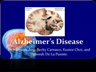
Brain Atrophy in AD WRONG! http://abdellab.sunderland.ac.uk/lectures/Neurodegeneration/References/Brain_Neurons_AD_Normal.html
AmyloidPlaques contain large amounts of a 42 amino acid peptide termed “b-amyloid”, or Ab42 b-amyloid itself is the initial cause of the pathophysiology that leads to dementia. Neurofibrillary tangles: rich in cytoskeletal proteins, especially the microtubule-associated protein, “tau”. In the tangles: heavily phosphorylated proteins, which may cause aggregation and precipitation of the cytoskeleton. The Anatomical Hallmark of Alzheimer’s Pathology: Amyloid Plaques and Neurofibrillary Tangles in Brain Also generally reduced brain volume, especially in entorhinal cortex and hippocampus From http://www.rnw.nl/health/html/brain.html
APP Protein: b a g g (2) g-secretase cuts this residue, giving: Ab40 Fragment or Soluble Ab42 Fragment Unsoluble,aggregates intoplaques b-secretase Pathway: (not drawn to scale) (1) b-secretase cuts APP protein, giving:
Amyloid precursor protein (APP) is membrane protein that sits in the membrane and extends outward. It is though to be important for neuronal growth, survival, and repair. From: www.niapublications.org/pubs/unraveling/01.htm
Enzymes cut the APP into fragments, the most important of which for AD is called b-amyloid (beta-amyloid) or Ab. From: www.niapublications.org/pubs/unraveling/01.htm
Beta-amyloid is “sticky” so the fragments cling together along with other material outside of the cell, forming theplaques seen in the AD brain. From: www.niapublications.org/pubs/unraveling/01.htm
Two Major Hypotheses for AD:b amyloid protein (BAP) v. tau • BAPtists: The accumulation of a fragment of the amyloid precursor protein or APP (the amyloid beta 42 residue fragment orAb-42) leads to the formation of plaques that kill neurons. • TAUists: Abnormal phosphorylation of tau proteins makes them “sticky,” leading to the break up of microtubules. The resultingloss of axonal transport causes cell death.
Amyloid Hypothesis(it’s the plaques) • The amyloid precursor protein (APP) is broken down by a series of secretases (see previous two slides). • During this process, a nonsoluble fragment of the APP protein (called Ab-42) accumulates and is deposited outside the cell. • The nonsoluble or “sticky” nature of Ab-42 helps other protein fragments(including apoE) to gather into plaques. • Somehow the plaques (or possible the migration of Ab-42 outside thecell) cause neuronal death. • PSEN1 & PSEN2 genes subunits of g secretase.
Tangles Axon Microtubules Neuron Tau Proteins Paired Helical Filament Dendrites Tau proteins, which normally stabilize microtubules in brain cells... thus creating tangles that disrupt cell functions and lead to cell death. undergo abnormal chemical changes and assemble into spirals called paired helical filaments... Theories of How DamageOccurs in AD From Inside the Cell: Tangle Formation Sources: Dr John Trojanowski and Dr Virginia M. Y. Lee. University of Pennsylvania Medical Center.
Microtubules are like railroad tracks that transport nutrition and other molecules. Tau-proteins act as “ties” that stabilize the structure of the microtubules. In AD, tau proteins become tangled, unstabilizing the structure of the microtubule. Loss of axonal transport results in cell death.
Loss of cholinergic neurons in the basal forebrain nuclei; therefore, restoring cholinergic function might be helpful. • Choline acetyltransferase (CAT) activity is reduced in cortex and hippocampus. • Nicotinic, but not muscarinic, receptors are reduced.
Therapeutic approaches • Cholinesterase inhibitors: • The first drugs approved for treating AD. • Enhancement of cholinergic transmission might compensate for the cholinergic deficit. • Improve cognitive performance. • Inhibiting excitotoxicity • Inhibiting neurodegeneration (future target).
Cholinesterase inhibitors • Tacrine • The first drug approved for treating AD. • Four times daily • Cholinergic side effects (nausea and abdominal cramps) • Hepatotoxicity
Cholinesterase inhibitors • Donepezil (no hepatotoxicity) • Rivastigmine: Long-lasting drug, CNS selective (fewer peripheral cholinergic side effects). • Galantamine: Acts partly by cholinesterase inhibitor and partly by activation of brain nicotinic AChRs.
Inhibiting excitotoxicity • Memantine • a use-dependent blockade of NMDA receptors.
Additional Treatments for AD • Role of dietary factors • Low saturated fat diets, • Vitamin E – decrease cytotoxicity - may slow the progression of the disease • Cholinergic stimulation: • Nicotine patch, varenicline (Chantix)
Pharmacologic Options for AD • Cognitive enhancers; can improve cognition and functional ability • 2 classes • Cholinesterase inhibitors (ChEIs) • NMDA-receptor antagonist
Common Side Effects National Institute on Aging. Alzheimer’s disease medications. November 2008. NIH PublicationNo. 08-3431. Available at: http://www.nia.nih.gov/Alzheimers/Publications/medicationsfs.htm. Accessed July 24, 2009.
Current theory: Multifactorial, involvingseveral pathways. • Protein accumulation: plaques & tangles • Inflammation: Unregulated activation of glia • Lipid distribution: Lipid membrane site of APP cleavage.
Targets for Future Therapies • A • -secretase inhibitors • -secretase inhibitors • Monoclonal antibodies • Tau protein • Inflammation • Insulin resistance
Inhibiting neurodegeneration (clinical trials) Phase II/ Phase III • Inhibitors of Ab aggregation (Immunological approach; antibody directed against Ab). • Inhibitors of b- and g-secretase. • Immunological approach (antibody directed against Ab). • Aβ vaccination • Statins: HMG-CaA reductase inhibitors
Inhibiting neurodegeneration (clinical trials) Phase II/ Phase III • Clioquinol (amoebicidal & metal chelating agent): Ab plaques bind cooper and zinc, thus removal of these metal ions promotes dissolution of the plaques. • NGF (Nerve growth factor): shortage of growth factors may contribute to the loss of forebrain cholinergic neurons in Alzheimer’s disease. • NSAIDs (esp. ibuprofen & indomethacin) reduce Ab42 formation by regulating g-secretase (unrelated to COX inhibition)