Download
1 / 35
350 likes | 490 Views
The Integumentary System. GPS Standard. SAP2. Students will analyze the interdependence of the integumentary, skeletal, and muscular systems as these relate to the protection, support and movement of the human body.

E N D
GPS Standard SAP2. Students will analyze the interdependence of the integumentary, skeletal, and muscular systems as these relate to the protection, support and movement of the human body. • Relate the structure of the integumentary system to its functional role in protecting the body and maintaining homeostasis.
Learning Goals • Describe the functions of the integumentary system. • Identify the structures of the skin and their functions. • Explain how the skin protects the body. • Explain how the skin maintains homeostasis. • List and describe the three pigments that contribute to skin color. • Describe how aging affects structures in the skin. • Describe two homeostatic imbalances of the skin.
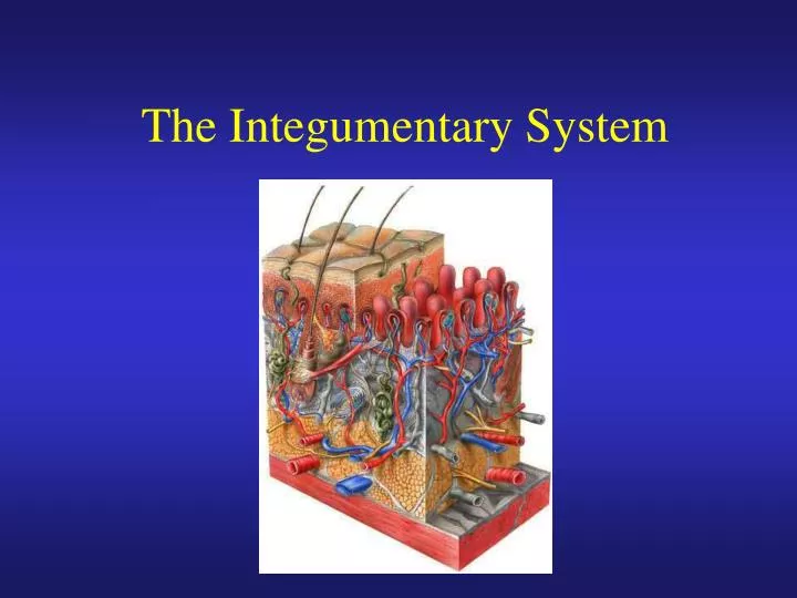
Introduction to the Integumentary System • The skin and its accessory structures make up the integumentary system. • The integumentary system functions to guard the body’s physical and biochemical integrity, maintain a constant body temperature, and provide sensory information about the surrounding environment. A large organ composed of all 4 tissue types 22 square feet 1-2 mm thick Weight 10 lbs.
TYPES OF SKIN • Thin skin • covers all parts of the body except for the palms and palmar surfaces of the digits and toes. • lacks epidermal ridges • has a sparser distribution of sensory receptors than thick skin. • Thick skin (0.6 to 4.5 mm) • covers the palms, palmar surfaces of the digits, and soles • features a stratum lucidum and thick epidermal ridges • lacks hair follicles, arrector pili muscles, and sebaceous glands, and has more sweat glands than thin skin.
FUNCTIONS OF SKIN • Thermoregulation • Perspiration & its evaporation • lowers body temperature • flow of blood in the dermis is adjusted • Shivering and constriction of surface vessels • raise internal body temperature as needed • Exercise • in moderate exercise, more blood brought to surface helps lower temperature • with extreme exercise, blood is shunted to muscles and body temperature rises
FUNCTIONS OF SKIN • blood reservoir • extensive network of blood vessels • protection - physical, chemical and biological barriers • tight cell junctions prevent bacterial invasion • lipids released retard evaporation • pigment protects somewhat against UV light • Langerhans cells alert immune system • cutaneous sensations • touch, pressure, vibration, tickle, heat, cold, and pain arise in the skin
FUNCTIONS OF SKIN • Synthesis of Vitamin D • activation of a precursor molecule in the skin by UV light • enzymes in the liver and kidneys modify the activated molecule to produce calcitriol, the most active form of vitamin D. • necessary vitamin for absorption of calcium from food in the gastrointestinal tract • excretion • 400 mL of water/day, small amounts salt, CO2, ammonia and urea
Cutaneous membrane Protects tissues from physical trauma, biological pathogens, and chemical trauma Provides sensations Accessory Structures Provides sensations Produces secretions Protects epidermal surfaces Integumentary System
Epidermis: Composed of keratinized stratified squamous epithelium Controls skin permeability Prevents entry of biological pathogens Synthesizes vitamin D Dermis: Composed of connective tissue and adipose Nourishes and supports epidermis Restricts spread of pathogens Stores lipids Attaches skin to underlying tissue Sensory receptors provide sensations Blood vessels assist in thermoregulation Cutaneous Membrane
Hair Follicles: Produce hair that protects underlying skin Provide sensations Nails: Protect and support the tips of fingers and toes Exocrine Glands: Assist in thermoregulation Excrete wastes Lubricate epidermis and hair Produce pheromones for chemical communication Accessory Structures
Specific Layer of the Epidermis Stratum Corneum: • Composed of 25 or more layers of dead squamous cells; shinglelike dead cell remnants completely filled with keratin Stratum Lucidum: • Observed only in non-hairy or thick skin. Several layers of clear, dead cells with indistinct boundaries
Specific Layers of the Epidermis Stratum Granulosum: Cells contain granules of keratin Stratum Spinosum: • Cells are pushed upward and flatten out • Appear spiny Stratum Basale: • Mitotic layer, continuously replaces epidermal cells, turnover rate between 25 to 50 days
Specialized Cells of the Epidermis Keratinocytes: Most common cells of the epidermis. Provides protection and waterproofing sealant Melanocytes: Produces and transfer the protein melanin to Keratinocytes. Melanin is a brown/black pigment that absorbs UV-light.
Glandular Epithelium • Gland: • a single cell or a mass of epithelial cells adapted for secretion • Endocrine glands are ductless. They secrete hormones into the bloodstream to help maintain homeostasis • Exocrine glands are connected to ducts that secrete---sweat, ear wax, saliva, digestive enzymes onto free surface of epithelial layer
Skin Color Pigments • Melanin produced in epidermis by melanocytes • UV in sunlight increases melanin production; • same number of melanocytes in everyone, but differing amounts of pigment produced; • results vary from yellow to tan to black color • Clinical observations • freckles or liver spots = melanocytes in a patch • albinism = inherited; no pigment • vitiligo = autoimmune loss of melanocytes in areas of the skin produces white patches • The wide variety of colors in skin is due to three pigments - melanin, carotene, and hemoglobin (in blood in capillaries) - in the dermis.
Skin Color Pigments • Carotene in dermis: • yellow-orange pigment (precursor of vitamin A) • found in stratum corneum & dermis • Hemoglobin in dermis: • red, oxygen-carrying pigment in blood cells • if other pigments are not present, epidermis is translucent so pinkness will be evident
Layers of the Dermis Dermis • Papillary Region: contain dermal papilla • Dermal Papilla: Fingerlike projections that contain a blood supply and pain receptors • Reticular region: deepest skin layer; contains blood vessels, sweat and oil glands, and deep pressure receptors; contain phagocytes that prevent bacteria from penetrating deeper into the body 2 1 3
Apocrine sweat glands: Found in the Axillae, nipples, labia, and glans penis. Begin to function at puberty and are affected by hormones Produce odorous thick secretion Possible pheromone function Sudoriferous (eccrine) Sweat Gland: Produce thin watery secretion Controlled by nervous system Thermoregulation Excretion of urea Antibacterial action Accessory Structures of the Dermis
Accessory Structures of the Dermis Sebaceous “Oil” gland: • Secrete sebum • Coats hair shaft and lubricates the epidermis • Secreted to hair follicles • Not association with hair on the labia, glans penis, and lips • Activity controlled by sex-hormones • Modified in external ear canal to produce cerumen or ear wax (ceruminous glands)
Special Sensory apparatus of the dermis Meissner’s Corpuscles: • Present in dermal papilla • Specialized sensory neuron nerve endings • Respond to touch • Most numerous in thick or non-hairy skin of the palmar and plantar surfaces
Special Sensory Apparatus of the Dermis Pacinian Corpuscle: • Encapsulated sensory nerve ending • Located at the hypodermis/dermis junction • Responds to pressure • Cross-section looks like an onion
Age Related Structural Changes • Collagen fibers decrease in number & stiffen (skin wrinkles, forms lines and folds) • Elastic fibers become less elastic (skin wrinkles) • Fibroblasts decrease in number (wounds slow to heal) • Decrease in number of melanocytes (gray hair, blotching) • Decrease in Langerhans cells (decreased immune responsiveness) • Reduced number and less-efficient phagocytes (less resistance to pathogens)
Resources • faculty.spokanefalls.edu/InetShare/.../Integumentary%20system.ppt