Download
1 / 9
90 likes | 271 Views
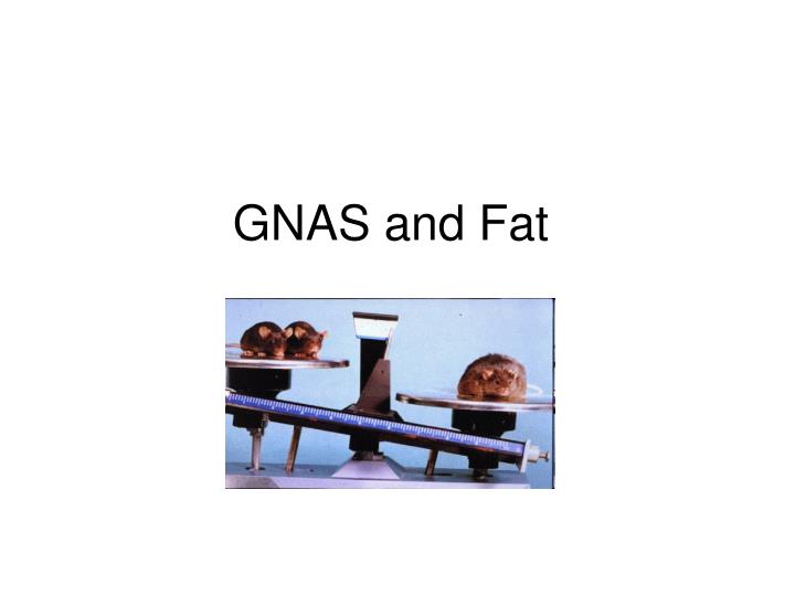
GNAS and Fat. KO. WT. White Adipose Tissue. KO. WT. WT. Brown adipose tissue. Fat Pads. WT KO. BAT. BAT. WAT. WAT. Observation: small fat pads in KO Possible reasons: Fewer adipocytes but same size Same number of adipocytes, but smaller (contain less lipid). (1). (2). KO.

E N D
KO WT White Adipose Tissue KO WT WT Brown adipose tissue Fat Pads
WT KO BAT BAT WAT WAT
Observation: small fat pads in KO • Possible reasons: • Fewer adipocytes but same size • Same number of adipocytes, but smaller (contain less lipid) (1) (2) KO WT KO WT
White Adipose Tissue • Yellow: lipid • Black: nuclei • Red: stroma • White: lipid • Light purple: stroma • Dark purple: nuclei • Black: lipid and nuclei • White: stroma Camera software Adobe Photoshop Conversion Image J Conversion
WAT Histomorphometry Parameters Percent stroma Percent adipocyte Total area Adipocyte number Adipocyte area Mean cell size Number of adipocytes/unit area
• Adipocytes smaller in KO • Number of adipocytes per unit area is greater • Area of fat/lipid per unit area is the same (68.7% in KO, 68.9% in WT) • Total weight of WAT is still greater in WT (0.87g in WT, 0.35g in KO) WT KO
BAT WT 12μm WT 6μm