Download
1 / 25
270 likes | 680 Views
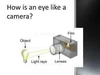
eye as a camera. KSJ Fig 27-3. optic disc. fovea. optic disc. Carpenter, Fig 26-1. demonstration of blind spot. photoreceptors in the retina. KSJ, Fig 26-1. retinal circuitry: laminar organization. KSJ, Fig 26-6. dynamic range of light intensity. Carpenter, Fig 7-3.

E N D
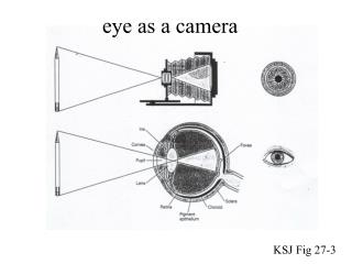
eye as a camera KSJ Fig 27-3
optic disc fovea optic disc Carpenter, Fig 26-1
photoreceptors in the retina KSJ, Fig 26-1
retinal circuitry: laminar organization KSJ, Fig 26-6
dynamic range of light intensity Carpenter, Fig 7-3
photopic vs scotopic vision photopic vision - at high light intensities - colour vision - high resolution - low sensitivity - best in fovea - Stiles-Crawford effect - mediated by cones scotopic vision - at low light intensities - achromatic - low resolution - high sensitivity - foveal scotoma - no Stiles-Crawford effect - mediated by rods
operating range: a sliding scale Carpenter, Fig 7.4
dark adaptation curves Sekuler and Blake, Fig 3-19
receptive fields of retinal ganglion cells KSJ, Fig 26-7
retina-LGN-cortex KSJ, Fig 27-4
LGN laminar organization KSJ Fig 27-6
LGN (and retinal) receptive fields achromatic colour-opponent KSJ, Fig 29-11
3 kinds of retinal ganglion cells parasol ("M") - 10 % - project to magnocellular layers of LGN - large dendritic fields, large fibres - large receptive fields -> low spatial frequencies, high velocities - achromatic midget ("P") - 80 % - project to parvocellular layers of LGN - small dendritic fields, small fibres - large receptive fields -> high spatial frequencies, low velocities - colour-opponent (red-green, possibly blue-yellow) bistratified (“K”) - 2 % - project to koniocellular layers of LGN - blue-yellow opponent
drifting grating stimuli: contrast contrast = (Lmax - Lmin) / (Lmax + Lmin) x 100% 100 % 50 % 25 % 12.5 % contrast sensitivity = 1 / contrast threshold
drifting grating stimuli: SF, TF, speed temporal frequency speed = ----------------------------- spatial frequency cycles/sec deg/sec = ---------------- cycles/deg
contrast sensitivity after M-lesions Merigan et al, Fig 2&3
effects of M vs P lesions: summary parvo lesion: - lower acuity - abolishes colour discrimination - reduced contrast sensitivity to gratings, at low temporal / high spatial frequencies (low velocities) magno lesion: - no effect on acuity - no effect on colour discrimination - reduced contrast sensitivity to gratings, at high temporal / low spatial frequencies (high velocities) - does not support idea of magno for motion, parvo for form vision
glaucoma: early detection central problem: need for early detection "at risk": ocular hypertension (OHT) perceptual "filling in" - example is failure to see your "blind spot" conventional (static) perimetry - detects problem only later human psychophysics, as approach for early detection: why you would not expect a deficit on many tasks: earliest lesions in peripheral vision, but many tasks use foveal vision -> need to do perimetry (automated) using the task task may be mediated by unaffected neurons, e.g. color-discrimination (P-cells)
Ganglion cell loss in glaucoma strategy #1: earliest effects on larger diameter fibres ( -> M-cells) theory: intra-ocular pressure block effects greatest on larger diameter fibers anatomy, in humans: fibre diameters, cell body sizes (Quigley et al) in animal models: experimentally raise IOP in monkeys (Dandona et al) 27 deg superior to fovea Quigley et al, Fig 11
motion coherence: stimulus see Adler’s, Fig 20-12, 22-11 task: report direction of motion noisy random dots: prevent using change-of-position a demanding task, requiring: combining responses of multiple neurons correct timing relations between neurons vary signal-to-noise (% coherence): best performance requires all the neurons
motion coherence: psychophysical thresholds % Correct Responses Motion Coherence (%)
motion coherence: loss in glaucoma Joffe et al (Fig 2)
selective M-cell loss hypothesis: criticisms apparent loss of large cells/fibres might be artifact of cell shrinkage also find losses of P-cell dependent psychophysics
testing for loss of sparse cell types strategy #2: most sensitive tests for capricious loss are those for sparse cell types: (explains loss of abilities that depend on M-cells) -> S-cones, blue/yellow (bistratified ganglion cells) color: detection of blue spot on yellow background rationale: blue-yellow ganglion cells (bistratified) are relatively sparse (ca 5%) results: Sample et al, Johnson et al: perimetry, longitudinal study