Download
1 / 69
820 likes | 1.94k Views
Thyroid diseases. Steven T. Nguy M.D. Assistant professor of Medicine Cooper University Hospitalist. TFT – A practical review. The hypothalamus-pituitary-thyroid function and thyroid physiology Usefulness and limitation of blood work studies in thyroid disorder Clinical cases. Hypothalamus.

E N D
Thyroid diseases Steven T. Nguy M.D. Assistant professor of Medicine Cooper University Hospitalist
TFT – A practical review • The hypothalamus-pituitary-thyroid function and thyroid physiology • Usefulness and limitation of blood work studies in thyroid disorder • Clinical cases
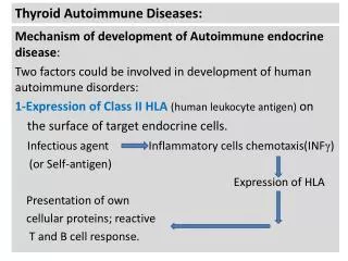
Hypothalamus TRH Pituitary Target Tissues Heart Thyroid Gland Liver T4 TR T3 Bone T4è T3 Liver CNS Hypothalamic-Pituitary-Thyroid AxisPhysiology – – TSH T4 T3 Adapted from Merck Manual of Medical Information. ed. R Berkow. 704:1997.
Thyroid Physiology • Thyroid gland releases T4 and T3 in molar ratio of 16:1 • >99% of the circulating thyroid hormones are bound to proteins (TBG, thyroxine-binding prealbumin, albumin) • The vast majority of circulating T3 is derived from deiodination of T4 outside the thyroid gland • Unbound (free) hormones T4 (0.03%) and T3 (0.3%) are biological active and are not influenced by TBG protein • In NTI, T4 conversion to T3 is reduced and conversion to rT3 is enhanced
Thyroid Funtion Tests • TSH • FT4, (T4) • T3, FT3 • Thyroglobulin • Thyroid stimulating immunoglobulin (TSI) or TSHR antibody • Antithyroid peroxidase antibodies (Anti TPO)
Serum TSH • Single best or initial test of the thyroid function. • Central to the negative-feedback system. • Inverse, log-linear relationship with thyroid hormone. Small changes in FT4 result in large changes in TSH level • Normal range 0.5 – 5.0 mU/L • Third generation TSH chemiluminometric assays have detection limits of about 0.01mU/L. • A normal TSH is sufficed to halt further testing unless suspect of possible hypothalamic pituitary disease.
Screening: Recommendations • Various societies and authors disagree about population-based screening • The USPSTF - insufficient evidences to recommend for or against routine screening for thyroid disease in adults. • The AAFP recommends screening high-risk populations: • women with a family hx of thyroid disease • women >35 y.o. • pregnant women • abnormal physical exam • diabetic patients • Hx of autoimmune disorder • The American Thyroid Association recommends screening start at age 35 (and q 5 years after that) Surks. JAMA. 2004 Jan 14;291(2):228-38. American Academy of Family Physicians. Subclinical Thyroid Disease. Available at: http://www.aafp.org/afp/20051015/1517.pdf Accessed February 16, 2006. The American Thyroid Association Web site. American Thyroid Association Guidelines for Detection of Thyroid Dysfunction. Available at: http://thyroid.org/professionals/publications/documents/GuidelinesdetectionThyDysfunc_2000.pdf. Accessed February 16, 2006.
Serum T4 • measured by radioimmunoassay (RIA), chemiluminometric assay, or similar immunometric technique. • 99.97% of serum T4 is bound to TBG (thyroxine binding globulin), transthyretin or TBPA (thyroxine-binding prealbumin), or albumin. • Serum total T4 assays measure both bound and unbound (“free”) T4 • Levels are high in approximately 90% of hyperthyroid patients and low in approximately 85% of hypothyroid patients.
Serum Free T4 • FT4 is measured by equilibrium dialysis techniques or estimated indirectly by calculation of free-thyroxine index (FTI) • FTI = T4 x T3 resin uptake (T3RU)% (T7, thyroid hormone-binding ratios) • FT4 assay is preferred test with tsh.
T3, Free T3, and rT3 • T3 - bindingprotein dependent. - Levels can be misleading in patients with acute illness, cirrhosis, uremia, or malnutrition. • FT3 - Useful to distinguish T3 toxicosis from subclinical thyrotoxicosis. • Reverse T3 (rT3) - increased in NTI. - inactive. - helpful to exclude central hypothyroidism
Other Ancillary Tests • Serum thyroglobulin – produced and released by thyroid gland. - marker for recurrent thyroid cancer - differentiate Graves disease from factitious thyrotoxicosis • Serum thyroid-stimulating immunoglobulin (TSI) or TSHR-Ab - Expensive test - Graves’ disease. • Antithyroid peroxidase antibodies (Anti TPO) – organ-specific and sensitive. - Hashimoto’s thyroiditis - predict overt hypothyroidism
Case 1 TSH 9.0 (0.5 – 5.5 mU/L) FT4 1.1 (0.8-1.8 ng/dL) What are your differential diagnoses?
Differential diagnoses for elevated TSH with normal FT4 TSH 9.0 (0.5-5.5 mU/L), T4 1.1 (0.8-1.8 ng/dL) • Transitional thyroid state or recovery from hypothyroidism (Hashimoto’s thyroiditis) • TSH receptor inactivating mutation • Patient with hypothyroidism who is on or recently started on treatment and the TSH hasn’t enough time to adjust. • Subclinical hypothyroidism
Case 1 A 75 yo woman with no prior medical problem presents for her annual physical examination. She says that she is slowing down a bit and has become more forgetful. She also mentions that she feels cold and is constipated. Physical examination is essentially within normal. Her blood work shows TSH 9.5 (0.5-4.5 mU/L) FT4 1.1 (0.8-1.8 ng/dL), normal C7, and CBCD. Which of the following actions would be most appropriate? • She should be treated with levothyroxine 0.1 mg/day. • Check lipid profile and start her on treatment if her HDL is low. • Check anti TPO level. If it shows high titer, she would be less likely become overt hypothyroidism in the future. • Repeat TSH and FT4 in 2 – 12 weeks.
Subclinical Thyroid DysfunctionClinical Guidelines By USPSTFAnn Intern Med. 2004;140:125-127,128-141JAMA, 2004;291:228-238,239-243 • Subclinical hypothyroidism is an elevation of serum TSH level with a FT4 in (low) normal range. • Upper limit of normal TSH should remain 4.5 or 5 mU/L • There was insufficient evidence linking subclinical hypothyroidism to systemic symptoms, cardiac dysfunction, adverse cardiac endpoints, LDL elevation and neuropsychiatric symptoms for TSH values ranging from 4.5 to 10 mU/L • The evidence for an association with dyslipidemia was rate as ‘‘fair’’, and only in individuals with TSH >10. • The decision to begin levothyroxine therapy in patient with persistent TSH but normal FT4 should be individualized. • If treatment is given, be cautious not to overtreat. Goal TSH 2.5-3.5
Other Ancillary Tests • Serum thyroglobulin – produced and released by thyroid gland. Level is elevated in conditions that increase thyroid gland function or destruction. It can be used as a tumor marker for recurrent of thyroid cancer or to differentiate Graves’disease from factitious thyrotoxicosis. • Serum thyroid-stimulating immunoglobulin (TSI) or TSHR-Ab – Expensive. Highly specific for Graves disease Generally not needed, but can be helpful in suspected cases of Graves’ disease in pregnancy or euthyroid ophthalmopathy. • Antithyroid peroxidase antibodies (Anti TPO) – useful in suspected case of Hashimoto’s thyroiditis, and in predicting for the development of overt hypothyroidism. It is highly organ-specific and sensitive.
Case 1 answer A 75 yo woman with no prior medical problem presents for her annual physical examination. She says that she is slowing down a bit and has become more forgetful. She also mentions that she feels cold and is constipated. Physical examination is essentially within normal. Her blood work shows TSH 9.0 (0.5-4.5 mU/L) FT4 1.1 (0.8-1.8 ng/dL), normal C7, and CBCD. Which of the following actions would be most appropriate? • She should be treated with levothyroxine 0.1 mg/day. • Check lipid profile and start her on treatment if her HDL is low. • Check anti TPO level. If it shows high titers, she would be less likely to become overt hypothyroidism in the near future • Repeat TSH and FT4 in 2 – 12 weeks.
Case 1 Answer • This patient has subclinical hypothyroidism. Her symptom is nonspecific. It is recommended to recheck TSH and FT4 in 2 – 12 weeks. If it is still elevated then consider start treatment with low dose levothyroxine and to recheck TSH again in 6 – 8 weeks. Goal is to get TSH down to normal range 2.5 – 3.5. The starting dose in choice A is too high and patient would likely become symptomatic from thyrotoxicosis. There is insufficient evidence to link subclinical hypothyroidism with elevated LDL for TSH 5.5 – 10. It has no effect on HDL. Patient with high titer anti TPO are more likely to become overt hypothyroidism (annualize rate is about 4.5%) but the current consensus didn’t advise the use of antiTPO antibodies in the decision making process.
Case 2 TSH 0.18 (0.45 – 5.0 mU/L) FT4 1.6 (0.8 – 1.8 ng/dL) What are your differential diagnoses?
Causes of low TSH and normal FT4 • Subclinical thyrotoxicosis (toxic multinodular goiter, toxic adenomas, mild Graves’disease, excessive replacement therapy) • Total triiodothyronine thyrotoxicosis (Early Graves’disease, autonomous thyroid nodules) • Drug effect (corticosteroids, octreotide, dopamine) • Non-thyroidal illness (euthyroid sick) • Transitional thyroid state (recovery from thyroiditis) • Excessive thyroid hormone therapy • Central hypothyroidism
Case 2 A 60 yo male with history htn, dm type 2, and hypercholesterolemia who has been doing well with medications including lisinopril 5mg daily, HCTZ 12.5 mg daily, glipizide XL 5 mg daily, and simvastatin 20 mg daily. Vital signs show T.98.5, pulse 100, resp rate 18, and pulsox 98% on room air. Phys exam is unremarkable. His routine lab shows TSH 0.18 (0.45 – 5.0 mU/L), FT4 1.6 (0.8 – 1.8 ng/dL), FT3 3.5 (1.5-7.0 pmol/L). The remaining of his labs including C7, CBCD, lipid profile, and HbA1C are within normal limit Which of the following statements would be appropriate? • He has subclinical hypothyroidism and very likely to develop osteosporosis and atrial fibrillation so he should be treated. • He should have a level of anti thyroid peroxidase antibody check. If it’s positive he will likely develop Graves’ disease in the future, so he should be treated. • He should have thyroid ultrasound, RAIU, and Echocardiogram. • He should have a repeat TSH and FT4 in 4 – 6 weeks • Start low dose PTU and recheck TSH and FT4 in 3 – 6 weeks
Subclinical Thyroid DysfunctionClinical Guidelines By USPSTFAnn Intern Med. 2004;140:125-127,128-141JAMA, 2004;291:228-238,239-243 • Subclinical hyperthyroidism is a suppressed serum TSH value resulting from an increase in serum T4 and/or T3 within the confines of the normal range • Evidence for adverse effects on the skeleton and the heart was “fair” to “good”, but only in patients with serum TSH levels < 0.1 mU/L • It is recommended that treatment “be considered” in patients with TSH levels < 0.1 mU/L even though there is a “paucity of intervention trials” showing benefit • For patients with the serum TSH levels between 0.1 and 0.45 mU/L, the evidence for adverse health consequences was insufficient or absent, and therefore therapy was not recommended.
Case 2 A 60 yo male with history htn, dm type 2, and hypercholesterolemia who has been doing well with medications including lisinopril 5mg daily, HCTZ 12.5 mg daily, glipizide XL 5 mg daily, and simvastatin 20 mg daily. Vital signs show T.98.5, pulse 100, resp rate 18, and pulsox 98% on room air. Phys exam is unremarkable. His routine lab shows TSH 0.18 (0.45 – 5.0 mU/L), FT4 1.6 (0.8 – 1.8 ng/dL), FT3 3.5 (1.5-7.0 pmol/L). The remaining of his labs including C7, CBCD, lipid profile, and HbA1C are within normal limit. EKG is also normal Which of the following statements would be appropriate? A. He has subclinical hypothyroidism and very likely to develop osteosporosis and atrial fibrillation so he should be treated. B. He should have a level of anti thyroid peroxidase antibody check. If it’s positive he will likely develop Graves’ disease in the future. C. He should have thyroid ultrasound, RAIU, and Echocardiogram. D. He should have a repeat TSH and FT4 in 3 – 6 weeks. E. Start low dose PTU and recheck TSH and FT4 in 3 – 6 weeks.
Case 2 Answer • This patient has subclinical hypertyroidism and is doing well. Current evidences suggest that risks of having osteosporosis and atrial fibrillation are “fair” to “good” but only in patients with TSH <0.1 mU/L. In patients with thyrotoxicosis, the presence of thyroglobulin-receptor antibodies, not anti TPO antibodies, in the serum is diagnostic of Graves’ disease, but the antibodies are absent in approximately 5 to 20 percent of patients with hyperthyroid Graves’ disease and almost certainly in a greater proportion of those with subclinical hyperthyroidism. Additional imaging or radioiodine uptake studies are not indicate as patient has no abnormal finding on exam. So at this time no treatment is needed and patient should have a repeat TFT in 4-6 weeks.
Case 3 A 60 year old woman with hx afib, HTN, and DM type 2 presents to ED complaining of feeling nervous and difficulty with sleep. She admits to have only mild palpitation but no CP or diaphoresis and the remaining of ROS are negative. Her medication includes amiodarone 200mg daily, metoprolol XL 50mg daily, metformin 500mg BID, and simvastatin 20mg bedtime. She appears mild anxious but no distress. VS. BP 134/78, T. 98.7, P.108, R.22 Neck exam shows slight tenderness and mild enlarged thyroid, heart exam is tachycardia with regular rhythm and a soft systolic murmur. The remaining of exam is within normal. Lab shows TSH is 0.01 (0.5-5.0 mU/L), FT4 50.3 (10.3-30.6 pmol/L). Remaining labs are within normal including C7, CBCD, CK, and TnT. Which of the following statements are true? • Patient has amiodarone induced thyroiditis so amiodarone must be stopped immediately and start on PTU or methimazole low dose. • Order color flow Doppler of thyroid gland, RAUI, and IL6 to determine the type of thyrotoxicosis • Treat patient with both a thionamide and prednisone, continue amiodarone, and recheck TSH and FT4 in 2-4 weeks • Stop amiodarone and recheck TSH, FT4 in 4-6 weeks. • She should be start on ASA and prednisone if the RAIU is high
Amiodarone induced thyrotoxicosis • About 3% of amiodarone-treated patients in the United States become hyperthyroid. (Hypothyroidism is more common than hyperthyroidism) • Two basic mechanisms in AIT Type I – Increase synthesis of T4 and T3 - Pre-existing multinodular goiter or latent Graves’ disease. More commonly seen in iodine-deficient areas of the world Type II – Direct toxic effect of amiodarone causing thyroiditis and hence release of T4 and T3 without increased hormone synthesis. More commonly seen in iodine-sufficient countries
Amiodarone induced thyrotoxicosis • Distinction between the two types is critical because the treatment is different. • Criteria used to attempt to distinguish type I from type II are 24-hour radioiodine uptake – if detectable, it suggest type I AIT Goiters – if has multinodular or diffuse goiter, it is more likely type I AIT. Serum thyroglobulin – higher in type I Serum IL-6 – higher in type II Color-flow Doppler sonography – may distinguish type I (increased vascularity) from type II (absent vascularity) hyperthyroidism.
Amiodarone induced thyrotoxicosis Should amiodarone be discontinued? • There are no good data that answer this question; however, the following should be considered: • Amiodarone may be necessary to control a life threatening arrythmia. • It has a very long half-life so stopping it would not give any immediate benefit. • Amiodarone appears ameliorate hyperthyroidism by blocking T4 to T3 conversion, beta-adrenergic receptors, and possibly T3 receptors. Stopping amiodarone might actually exacerbate hyperthyroid symptoms and signs.
Amiodarone induced thyrotoxicosis treatment • Type I AIT . Drugs-Thionamide (PTU or methimazole) is the first line therapy (whether amiodarone is continued or discontinued). Higher than average doses are often needed . Radioiodine ablation – if the RAIU is high enough. . Surgery – only if refractory to antithyroid drug therapy. • Type II AIT . Glucocorticoids – Prednisone 40-60 mg/day. Continue therapy for one to two months before tapering • “Mixed” type I and type II AIT . Combination of glucocorticoid and thionamine initially. A rapid response suggests type II, the thionamide can then be tapered or stopped. A poor or slow initial response argues for type I AIT
Case 3 A 60 year old woman with hx afib, HTN, and DM type 2 presents to ED complaining of feeling nervous and difficulty with sleep. She admits to have only mild palpitation but no CP or diaphoresis and the remaining of ROS are negative. Her medication includes amiodarone 200mg daily, metoprolol XL 50mg daily, metformin 500mg BID, and simvastatin 20mg bedtime. She Appears mild anxious but no distress. VS. BP 134/78, T. 98.7, P.108, R.22 Neck exam shows slight tenderness and mild enlarged thyroid, heart exam is tachycardia with regular rhythm and a soft systolic murmur. The remaining of exam is within normal. Lab shows TSH is 0.01 (0.5-5.0 mU/L), FT4 50.3 (10.3-30.6 pmol/L). Remaining labs are within normal including C7, CBCD, CK, and TnT. Which of the following statements are true? • Patient has amiodarone induced thyroditis so amiodarone must be stopped immediately and start on PTU or methimazole low dose. • The color flow Doppler of thyroid gland, RAUI, and IL6 may help to determine the type of thyrotoxicosis • Treat patient with both a thionamide and prednisone, continue amiodarone, and recheck TSH and FT4 in 2-4 weeks • Stop amiodarone and recheck TSH, FT4 in 4-6 weeks. • She should be start on ASA and prednisone if the RAIU is high
Case 3 answer B and C are the correct statement. A is false because amiodarone has a very long half-life so stopping it would not have any immediate benefit and potentially can cause arrhythmia. D is false because patient needs treatment for thyrotoxicosis E is false because in acute thyrotoxicosis state aspirin can exacerbate the condition because it binds to TBG and causing more available unbound thyroxine.
Case 4 A 70 yo male patient was admitted to ICU 3 days ago for pneumonia, COPD exacerbation which required intubation. He was successfully extubated and transferred to telemetry floor yesterday. Overnight the telemetry shows sinus rhythm 80 to sinus tachycardia 105 with few atrial ectopy and a normal EKG. He is on Levaquin 750mg daily, duoneb Q4H, and hydrocortisone 60mg Q6H. He appears frail, weak and complains only of no appetite. The BP 98/70 T.99, P.100, RR. 20, pulsox 96% on 2L. On exam, he has RLL rhonchi but no crackles, heart rate is slightly fast but no murmur or rub. The remaining of his exam was unremarkable. AM lab shows WBC 13.0 Hb 12 Plt 200K, band 6%, seg. neutrophil 80%, normal C7, TSH 0.15 (0.45-4.5), T4 normal and T3 low. Which of the following would be appropriate to do next? • This patient has lab result suggestive of central hypothyroidism so MRI of the head should be done first. • Order a baseline cortisol level and do a cosyntropin test to rule out adrenal insufficiency. • Order a serum rT3 level and if the level is high no other test is necessary. • Start patient on levothyroxine 0.025mg daily for hypothyroidism
Nonthyroidal illness (Euthyroid Sick Syndrome) • Abnormal findings on TFT that occur in the setting of a NTI without preexisting hypothalamic-pituitary and thyroid gland dysfunction. • The most prominent alterations are low serum T3 and elevated reverse T3 (rT3). • Serum TSH, T4, and FT4 are also affected in variable degrees based on the severity and duration of the NTI. • Probable mechanism: - Decreased or inhibition of 5’-monodeiodination (endogenous cortisol or exogenous glucocorticoid therapy, non-esterified fatty acids, cytokines TNF, IF, IL6.) - The peripheral production of T3 is decrease, but its clearance is unchanged; whereas, the production of rT3 is unchanged, while its clearance is diminished • Treatment is not needed. After recovery from an NTI, these thyroid function test result abnormalities should be completely reversible
Case 4 A 70 yo male patient was admitted to ICU 3 days ago for pneumonia, COPD exacerbation which required intubation. He was successfully extubated and transferred to telemetry floor yesterday. Overnight the telemetry shows sinus rhythm 80 to sinus tachycardia 105 with few atrial ectopy and a normal EKG. He is on Levaquin 750mg daily, duoneb Q4H, and hydrocortisone 60mg Q6H. He appears frail, weak and complains only of no appetite. The BP 98/70 T.99, P.100, RR. 20, pulsox 96% on 2L. On exam, he has RLL rhonchi but no crackles, heart rate is slightly fast but no murmur or rub. The remaining of his exam was unremarkable. AM lab shows WBC 13.0 Hb 12 Plt 200K, band 6%, seg. neutrophil 80%, normal C7, TSH 0.15 (0.45-4.5), T4 normal and T3 low. Which of the following would be appropriate to do next? • This patient has lab result suggestive of central hypothyroidism so MRI of the head should be done first. • Order a baseline cortisol level and do a cosyntropin test to rule out adrenal insufficiency. • Order a serum rT3 level and if the level is high no other test is necessary. • Start patient on levothyroxine 0.025mg daily for hypothyroidism
Case 4 answer • This patient has low TSH, low T3 and normal T4. The differential diagnoses includes central hypothyroidism, euthyroid sick syndrome (NTI), or patient with hyperthyroidism undergoing treatment with antithyroid medication. Base on the information given (the patient recently went through physiological stressful event, and is on corticosteroid and no history of hyperthyroidism) the patient most likely has euthyroid sick syndrome. In NTI the activities of 5’-monodeiodinase is decreased or inhibited so the peripheral conversion of T3 diminishes and the clearance of rT3 is reduced causing low serum T3 and high rT3. There are no abnormal finding on neuro exam and so MRI would not be the first test. Cotrosyn test on this patient would be inappropriate because he is on exogenous glucocorticoid therapy (except dexamethasone) which interfere with the test and aside from that his C7 is normal so adrenal insufficiency is unlikely. In NTI no treatment is necessary.
Case 5 A 28 year-old woman presents with a palpable mass on the left side of her neck. She has no neck pain and no symptoms of thyroid dysfunction. Physical exam reveals a solitary, mobile thyroid nodule, 2 x 3 cm, without lymphadenopathy. The patient has no family history of thyroid disease and no history of external radiation. A blood drawn was sent for serum TSH and FT4. Which of the following statements are true? • If TSH is low and FT4 high, she has hyperthyroidism so no further evaluation needed and start treatment with antithyroid medication. • If TSH is elevated check anti TPO antibody level. If it is elevated she has Hashimotos thyroiditis and no further testing necessary and start treatment with thyroid medication. • Serum calcitonin level should be routinely check in young patient with thyroid nodule(s). • She would need to have thyroid ultrasound and scintigraphy regardless of the TSH level. • If TSH is normal, the next step to evaluate her nodule would be doing a FNAB (with ultrasound guidance.)
Thyroid Nodules • Palpable nodules occur in 4-7% of the population. • Studies suggest about 30% of subjects 19 to 50 years of age had an incidental nodule on ultrasonography. • Types of nodules Colloid, cysts, and thyroiditis (80%) Benign follicular neoplasms (10-15%) Thyroid carcinoma (5%) • History and physical exam remain the diagnostic cornerstones in evaluating the patient with thyroid nodule and may be suggestive of thyroid carcinoma
Algorithm for the Cost-Effective Evaluation and Treatment of a Clinically Detectable Solitary Thyroid Nodule Hegedus L. N Engl J Med 2004;351:1764-1771
Case 5 A 28 year-old woman presents with a palpable mass on the left side of her neck. She has no neck pain and no symptoms of thyroid dysfunction. Physical exam reveals a solitary, mobile thyroid nodule, 2 x 3 cm, without lymphadenopathy. The patient has no family history of thyroid disease and no history of external radiation. A blood drawn was sent for serum TSH and FT4. Which of the following statements is true? • If TSH is low and FT4 high, she has hyperthyroidism so no further evaluation needed and start treatment with antithyroid medication. • If TSH is elevated check anti TPO antibody level. If it is elevated she has Hashimoto’s thyroiditis and no further testing necessary and start treatment with thyroid medication. • Serum calcitonin level should be routinely check in young patient with thyroid nodule(s). • She would need to have thyroid ultrasound and scintigraphy regardless of the TSH level. • If TSH is normal, the next step to evaluate her nodule would be doing a FNAB (with ultrasound guidance.)
Case 5 answer • A is false because eventhough she has thyrotoxicosis one needs to check whether the nodule is hot or cold. A hot nodule is nearly always benign, whereas a nonfunctioning nodule, constituting approximately 90% of nodules, has a 5% risk of being malignant. Thus, in patient with a suppressed TSH and a hot nodule no further evaluation is necessary • B is false because although the elevated anti TPO level confirm Hashimoto’s thyroiditis one must rule out a coexisting cancer, including lymphoma, which accounts for only 5% of thyroid cancers but is associated with Hashimoto’s thyroiditis. • C is false because medullary carcinoma is rare (about 1 of 250 patients with thyroid nodule), it should be check only in patient with family history of MEN or medullary thyroid carcinoma • D is false because if she has normal TSH FNAB (with ultrasound guidance) would be the next step of evaluation. • E is the correct statement.
Case 6 A 30 yo woman presents to clinic with a complain of insomnia. She recently gave birth to a healthy baby boy 3 months ago and is still breastfeeding. ROS is also positive for nervousness, heat intolerance, and weight loss (she now weighs 5 lbs lighter than before her pregnancy) On exam she appears anxious, pulse 104, bp 140/68. Eye has lid lag but no proptosis or soft tissue inflammation. Neck has nontender thyroid gland twice the normal size, without bruit or nodules. She has fine tremor and moist palmar skin. DTR are brisk. Lab studies: serum FT4 49.7 (10.3-30.6pmol/L), serum TSH <0.01 (0.5-5.0 mU/L), normal C7, CBCD, and LFT. Which of the following statement would be done next? • Check EKG, UDAS, and RAIU. • Check anti TPO antibody. • Start on B blocker for post partum thyroiditis • Check serum anti TSI (thyroid-stimulating immunoglobulin) or anti TSHR.. • Start on SSRI for post partum depression.
Case 6 Answer Two common form of thyrotoxicosis developed shortly after pregnancy (1-4 months) are postpartum thyroiditis and Graves’ disease. It’s important to determine the cause of thyrotoxicosis in post partum because the course of treatment would be different. RAIU would be helpful to differentiate hyperthyroidism (Graves’ disease, autonomous thyroid nodules, and toxic mutlinodular goiter) from thyroiditis (subacute thyroiditis, postpartum thyroiditis) or exogenous thyroid hormone but it’s contraindicated in breasfeeding patient. Both postpartum thyroiditis and Graves’ disease are considered autoimmune diseases so a positive anti TPO antibody testing is not helpful. Serum TSI is more specific and is diagnostic for Graves’ disease. The treatment for Graves’ disease in postpartum include antithyroid drugs, radioactive iodine (if patient is not breastfeeding), and surgery. Postpartum thyroiditis is treated with observation, with or without B-Blocker during the thyrotoxic stage. Caution is indicated, however, because B-Blockers are secreted into breast milk. Starting SSRI treatment for depression or anxiety is inappropriate at this time because this patient has true abnormal thyroid function studies and medication may interfere with her alertness to care for an infant.
Case 7 An elderly woman comes to your clinic complaining of weakness, fatigue, and having no interest in life. On questioning, she reports cold intolerance, one bowel movement every 4-5 days, and some hair loss, but denies any weight gain. She is depressed and doesn’t care about herself or her home. BP 98/62 and HR 57 Labs show Na 140, K 5.2, Cl 109, HCO3 18, BUN 9, Cr 0.9, Glucose 70, FT4 0.3 (0.8-1.5ng/dL), and TSH 24.5 (0.5 – 5.0mU/L) What should you do next? • Begin oral thyroxine 100 mcg daily • Give one dose of IV thyroxine 500 mcg in One Day Stay and begin oral thyroxine 150 mcg daily • Begin oral thyroxine 300 mcg daily • Begin oral thyroxine 50 mcg daily • Begin oral dexamethasone 0.5 mg q a.m. first then oral thyroxine 100 mcg per day and perform an ACTH (cosyntropin) stimulation test
Case 7 Answer E is the correct answer – This patient has both hypothyroidism and adrenal insufficiency (Schmidt’s syndrome) Of course the thyroid function test confirm hypothyroidism but don’t miss other possible issue. She has several clues that raise the possibility of adrenal insufficiency: not gaining weight despite being hypothyroid, low blood pressure, and a hint of hyperkalemic metabolic acidosis. The blood pressure is not unusual for a woman, but hypothyroidism usually raises diastolic blood pressure. The bp alone is not diagnostic of adrenal insufficiency, but it highly suggestive when taken in context with other findings. The glucose is also low normal. It is imperative to recognize a possible case of Schmidt’s syndrome because the patients die soon after starting thyroid hormone replacement unless they begin glucocorticoid replacement. If you ever suspect that a patient may have adrenal insufficiency, you must begin steroids and work up the patient. The standard test is an ACTH1-24 (cosyntropin) stimulation test. Because dexamethasone is the only available glucocorticoid that doesn’t interfere with cortisol assay, the patient must be started on dexamethasone until the results of the tests are available. If she truly has adrenal insufficiency, it should be treated with hydrocortisone because it also has some mineralcorticoid activity. Once she takes her first steroid pill, she can begin thyroxine.
Case 8 A 60 year-old woman with hx multinodular goiter and CAD presents to hospital with c/o palpitation and nervous. She is found to have atrial fibrillation with a rapid ventricular response. One month ago when she was admitted for anginal chest pain and had an abnormal stress test. A follow up coronary arteriogram showed non-significant coronary artery disease. The thyroid function test at that time was within normal limit (TSH 0.8 mU/L). Thyroid function testing is repeated TSH <0.01 mU/L (0.5-4.5 mU/L) FT4 40.5 pmol/L (10.3-30.6 pmol/L) What is the most likely diagnosis? • Graves’ disease • Stress-induced hyperthyroidism • Iodine-induced hyperthyroidism • Silent thyroiditis • Euthyroid sick syndrome